 Movie
Movie Controller

[English] 日本語
 Yorodumi
- PDB-5o8i: Crystal structure of human histidine triad nucleotide-binding pro... -
+ Open data

- Basic information
| Entry | Database: PDB / ID: 5o8i | ||||||
|---|---|---|---|---|---|---|---|
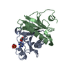
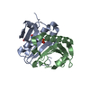
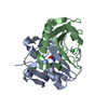
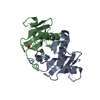
| Title | Crystal structure of human histidine triad nucleotide-binding protein 1 (hHINT1) crystallized at P212121 space group, and refined to 1.27 A | ||||||
 Components Components | Histidine triad nucleotide-binding protein 1 | ||||||
 Keywords Keywords | HYDROLASE / phosphoramidase / desulfurase / tumour suppressor | ||||||
| Function / homology |  Function and homology information Function and homology informationpurine ribonucleotide catabolic process / Hydrolases; Acting on phosphorus-nitrogen bonds / adenosine 5'-monophosphoramidase activity / deSUMOylase activity / protein desumoylation / Regulation of MITF-M-dependent genes involved in apoptosis / histone deacetylase complex / intrinsic apoptotic signaling pathway by p53 class mediator / Regulation of MITF-M-dependent genes involved in cell cycle and proliferation / Transcriptional and post-translational regulation of MITF-M expression and activity ...purine ribonucleotide catabolic process / Hydrolases; Acting on phosphorus-nitrogen bonds / adenosine 5'-monophosphoramidase activity / deSUMOylase activity / protein desumoylation / Regulation of MITF-M-dependent genes involved in apoptosis / histone deacetylase complex / intrinsic apoptotic signaling pathway by p53 class mediator / Regulation of MITF-M-dependent genes involved in cell cycle and proliferation / Transcriptional and post-translational regulation of MITF-M expression and activity / positive regulation of calcium-mediated signaling / protein kinase C binding / cytoskeleton / Hydrolases; Acting on peptide bonds (peptidases); Cysteine endopeptidases / nucleotide binding / hydrolase activity / regulation of DNA-templated transcription / signal transduction / proteolysis / extracellular exosome / nucleoplasm / nucleus / plasma membrane / cytoplasm / cytosol Similarity search - Function | ||||||
| Biological species |  Homo sapiens (human) Homo sapiens (human) | ||||||
| Method |  X-RAY DIFFRACTION / SYNCHROTRON / MOLECULAR REPLACEMENT / Resolution: 1.27 Å X-RAY DIFFRACTION / SYNCHROTRON / MOLECULAR REPLACEMENT / Resolution: 1.27 Å | ||||||
 Authors Authors | Dolot, R.M. / Seda, A. / Nawrot, B.C. | ||||||
 Citation Citation | Journal: To Be Published Title: Differences in crystal packing as the key factor for stabilization of the N-terminal fragment of hHINT1 protein Authors: Dolot, R.M. / Seda, A. / Nawrot, B.C. | ||||||
| History |
|
- Structure visualization
| Structure viewer | Molecule: MolmilJmol/JSmol |
|---|
- Downloads & links
-Download
| PDBx/mmCIF format | 5o8i.cif.gz | 133.9 KB | Display | PDBx/mmCIF format |
|---|---|---|---|---|
| PDB format | pdb5o8i.ent.gz | 103.6 KB | Display | PDB format |
| PDBx/mmJSON format | 5o8i.json.gz | Tree view | PDBx/mmJSON format | |
| Others |  Other downloads Other downloads |
-Validation report
| Arichive directory | https://data.pdbj.org/pub/pdb/validation_reports/o8/5o8iftp://data.pdbj.org/pub/pdb/validation_reports/o8/5o8i | HTTPS FTP |
|---|
-Related structure data
| Related structure data |  6g9zC 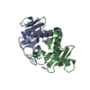 3tw2S S: Starting model for refinement C: citing same article ( |
|---|---|
| Similar structure data |


-Links
 PDBj
PDBj


- Assembly
| Deposited unit | 
| ||||||||||||||||||
|---|---|---|---|---|---|---|---|---|---|---|---|---|---|---|---|---|---|---|---|
| 1 |
| ||||||||||||||||||
| Unit cell |
| ||||||||||||||||||
| Noncrystallographic symmetry (NCS) | NCS domain:
NCS domain segments: Component-ID: _ / Ens-ID: 1 / Beg auth comp-ID: ARG / Beg label comp-ID: ARG / End auth comp-ID: PRO / End label comp-ID: PRO / Refine code: _ / Auth seq-ID: 12 - 125 / Label seq-ID: 12 - 125
|
-Components
| #1: Protein | Mass: 13823.931 Da / Num. of mol.: 2 Source method: isolated from a genetically manipulated source Details: First eleven amino acid residues are not visible on electron density maps Source: (gene. exp.) Homo sapiens (human) / Gene: HINT1, HINT, PKCI1, PRKCNH1 / Plasmid: pSGA02 / Production host:  #2: Chemical | ChemComp-ADP / |   Mass: 427.201 Da / Num. of mol.: 1 / Source method: obtained synthetically / Formula: C10H15N5O10P2 / Comment: ADP, energy-carrying molecule*YM Mass: 427.201 Da / Num. of mol.: 1 / Source method: obtained synthetically / Formula: C10H15N5O10P2 / Comment: ADP, energy-carrying molecule*YM#3: Water | ChemComp-HOH / |  Mass: 18.015 Da / Num. of mol.: 414 / Source method: isolated from a natural source / Formula: H2O Mass: 18.015 Da / Num. of mol.: 414 / Source method: isolated from a natural source / Formula: H2O |
|---|
-Experimental details
-Experiment
| Experiment | Method: X-RAY DIFFRACTION / Number of used crystals: 1 |
|---|
- Sample preparation
| Crystal | Density Matthews: 2.58 Å3/Da / Density % sol: 52.35 % / Description: needles |
|---|---|
| Crystal grow | Temperature: 281 K / Method: vapor diffusion, hanging drop / pH: 8.5 Details: 20% w/v PEG 3350, 0.1 M Bis-Tris Propane pH 8.5, 0.2 M Sodium/Potassium Phosphate |
-Data collection
| Diffraction | Mean temperature: 100 K |
|---|---|
| Diffraction source | Source: SYNCHROTRON / Site: BESSY  / Beamline: 14.1 / Wavelength: 0.918409 Å / Beamline: 14.1 / Wavelength: 0.918409 Å |
| Detector | Type: DECTRIS PILATUS 6M / Detector: PIXEL / Date: Apr 30, 2016 |
| Radiation | Monochromator: Si(111) crystal / Protocol: SINGLE WAVELENGTH / Monochromatic (M) / Laue (L): M / Scattering type: x-ray |
| Radiation wavelength | Wavelength: 0.918409 Å / Relative weight: 1 |
| Reflection | Resolution: 1.27→40.43 Å / Num. obs: 74711 / % possible obs: 98.6 % / Redundancy: 4.1 % / Biso Wilson estimate: 8.4 Å2 / CC1/2: 0.999 / Rmerge(I) obs: 0.044 / Rpim(I) all: 0.038 / Net I/σ(I): 14.1 |
| Reflection shell | Resolution: 1.27→1.29 Å / Redundancy: 3.8 % / Rmerge(I) obs: 0.535 / Num. unique obs: 3586 / CC1/2: 0.775 / Rpim(I) all: 0.461 / % possible all: 95.8 |
- Processing
| Software |
| ||||||||||||||||||||||||||||||||||||||||||||||||||||||||||||||||||||||||||||||||||||||||||||||||||||||||||||||||||||||||||||||||||||||||||||||||||||||||||||||||||||||||||||||||||||||
|---|---|---|---|---|---|---|---|---|---|---|---|---|---|---|---|---|---|---|---|---|---|---|---|---|---|---|---|---|---|---|---|---|---|---|---|---|---|---|---|---|---|---|---|---|---|---|---|---|---|---|---|---|---|---|---|---|---|---|---|---|---|---|---|---|---|---|---|---|---|---|---|---|---|---|---|---|---|---|---|---|---|---|---|---|---|---|---|---|---|---|---|---|---|---|---|---|---|---|---|---|---|---|---|---|---|---|---|---|---|---|---|---|---|---|---|---|---|---|---|---|---|---|---|---|---|---|---|---|---|---|---|---|---|---|---|---|---|---|---|---|---|---|---|---|---|---|---|---|---|---|---|---|---|---|---|---|---|---|---|---|---|---|---|---|---|---|---|---|---|---|---|---|---|---|---|---|---|---|---|---|---|---|---|
| Refinement | Method to determine structure: MOLECULAR REPLACEMENT Starting model: 3TW2 Resolution: 1.27→40.43 Å / Cor.coef. Fo:Fc: 0.987 / Cor.coef. Fo:Fc free: 0.979 / SU B: 1.113 / SU ML: 0.021 / Cross valid method: THROUGHOUT / ESU R: 0.029 / ESU R Free: 0.032 / Details: HYDROGENS HAVE BEEN ADDED IN THE RIDING POSITIONS
| ||||||||||||||||||||||||||||||||||||||||||||||||||||||||||||||||||||||||||||||||||||||||||||||||||||||||||||||||||||||||||||||||||||||||||||||||||||||||||||||||||||||||||||||||||||||
| Solvent computation | Ion probe radii: 0.8 Å / Shrinkage radii: 0.8 Å / VDW probe radii: 1.2 Å | ||||||||||||||||||||||||||||||||||||||||||||||||||||||||||||||||||||||||||||||||||||||||||||||||||||||||||||||||||||||||||||||||||||||||||||||||||||||||||||||||||||||||||||||||||||||
| Displacement parameters | Biso mean: 16.25 Å2
| ||||||||||||||||||||||||||||||||||||||||||||||||||||||||||||||||||||||||||||||||||||||||||||||||||||||||||||||||||||||||||||||||||||||||||||||||||||||||||||||||||||||||||||||||||||||
| Refinement step | Cycle: 1 / Resolution: 1.27→40.43 Å
| ||||||||||||||||||||||||||||||||||||||||||||||||||||||||||||||||||||||||||||||||||||||||||||||||||||||||||||||||||||||||||||||||||||||||||||||||||||||||||||||||||||||||||||||||||||||
| Refine LS restraints |
|