 Movie
Movie Controller

[English] 日本語
 Yorodumi
- PDB-6g49: Crystal structure of the periplasmic domain of TgpA from Pseudomo... -
+ Open data

- Basic information
| Entry | Database: PDB / ID: 6g49 | ||||||
|---|---|---|---|---|---|---|---|
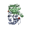
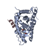
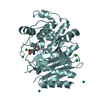
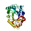
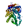
| Title | Crystal structure of the periplasmic domain of TgpA from Pseudomonas aeruginosa | ||||||
 Components Components | Protein-glutamine gamma-glutamyltransferase | ||||||
 Keywords Keywords | TRANSFERASE / essential bacterial protein | ||||||
| Function / homology |  Function and homology information Function and homology informationprotein-glutamine gamma-glutamyltransferase / protein-glutamine gamma-glutamyltransferase activity / plasma membrane Similarity search - Function | ||||||
| Biological species |  Pseudomonas aeruginosa PAO1 (bacteria) Pseudomonas aeruginosa PAO1 (bacteria) | ||||||
| Method |  X-RAY DIFFRACTION / SYNCHROTRON / MOLECULAR REPLACEMENT / Resolution: 1.6 Å X-RAY DIFFRACTION / SYNCHROTRON / MOLECULAR REPLACEMENT / Resolution: 1.6 Å | ||||||
 Authors Authors | Milani, M. / Mastrangelo, E. / Uruburu, M. | ||||||
 Citation Citation | Journal: J.Struct.Biol. / Year: 2019 Title: Structural and functional characterization of TgpA, a critical protein for the viability of Pseudomonas aeruginosa. Authors: Uruburu, M. / Mastrangelo, E. / Bolognesi, M. / Ferrara, S. / Bertoni, G. / Milani, M. | ||||||
| History |
|
- Structure visualization
| Structure viewer | Molecule: MolmilJmol/JSmol |
|---|
- Downloads & links
-Download
| PDBx/mmCIF format | 6g49.cif.gz | 85.9 KB | Display | PDBx/mmCIF format |
|---|---|---|---|---|
| PDB format | pdb6g49.ent.gz | 62.7 KB | Display | PDB format |
| PDBx/mmJSON format | 6g49.json.gz | Tree view | PDBx/mmJSON format | |
| Others |  Other downloads Other downloads |
-Validation report
| Summary document | 6g49_validation.pdf.gz | 449.4 KB | Display | wwPDB validaton report |
|---|---|---|---|---|
| Full document | 6g49_full_validation.pdf.gz | 454.8 KB | Display | |
| Data in XML | 6g49_validation.xml.gz | 17.4 KB | Display | |
| Data in CIF | 6g49_validation.cif.gz | 26.1 KB | Display | |
| Arichive directory | https://data.pdbj.org/pub/pdb/validation_reports/g4/6g49ftp://data.pdbj.org/pub/pdb/validation_reports/g4/6g49 | HTTPS FTP |
-Related structure data


-Links
 PDBj
PDBj
- Assembly
| Deposited unit | 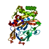
| ||||||||
|---|---|---|---|---|---|---|---|---|---|
| 1 |
| ||||||||
| Unit cell |
|
-Components
| #1: Protein | Mass: 38442.125 Da / Num. of mol.: 1 Source method: isolated from a genetically manipulated source Details: protein TAG: MGSDKIHHHHHHHHHHGV / Source: (gene. exp.) Pseudomonas aeruginosa PAO1 / Gene: tgpA, PA2873 / Production host: References: UniProt: Q9HZX3, protein-glutamine gamma-glutamyltransferase | ||||
|---|---|---|---|---|---|
| #2: Chemical | ChemComp-PO4 /   Mass: 94.971 Da / Num. of mol.: 7 / Source method: obtained synthetically / Formula: PO4 Mass: 94.971 Da / Num. of mol.: 7 / Source method: obtained synthetically / Formula: PO4#3: Chemical | ChemComp-CL / |   Mass: 35.453 Da / Num. of mol.: 1 / Source method: obtained synthetically / Formula: Cl Mass: 35.453 Da / Num. of mol.: 1 / Source method: obtained synthetically / Formula: Cl#4: Water | ChemComp-HOH / |  Mass: 18.015 Da / Num. of mol.: 287 / Source method: isolated from a natural source / Formula: H2O Mass: 18.015 Da / Num. of mol.: 287 / Source method: isolated from a natural source / Formula: H2O |
-Experimental details
-Experiment
| Experiment | Method: X-RAY DIFFRACTION / Number of used crystals: 1 |
|---|
- Sample preparation
| Crystal | Density Matthews: 2.74 Å3/Da / Density % sol: 55.13 % |
|---|---|
| Crystal grow | Temperature: 293 K / Method: batch mode / pH: 7.5 Details: 0.1 M HEPES sodium pH 7.5 0.8 M Sodium phosphate monobasic monohydrate 0.8 M Potassium phosphate monobasic |
-Data collection
| Diffraction | Mean temperature: 100 K |
|---|---|
| Diffraction source | Source: SYNCHROTRON / Site: ESRF  / Beamline: MASSIF-3 / Wavelength: 0.9677 Å / Beamline: MASSIF-3 / Wavelength: 0.9677 Å |
| Detector | Type: DECTRIS EIGER R 4M / Detector: PIXEL / Date: Apr 1, 2016 |
| Radiation | Protocol: SINGLE WAVELENGTH / Monochromatic (M) / Laue (L): M / Scattering type: x-ray |
| Radiation wavelength | Wavelength: 0.9677 Å / Relative weight: 1 |
| Reflection | Resolution: 1.6→43.5 Å / Num. all: 429906 / Num. obs: 57370 / % possible obs: 99.9 % / Redundancy: 7.5 % / CC1/2: 0.999 / Rpim(I) all: 0.032 / Rrim(I) all: 0.064 / Net I/σ(I): 17.6 |
| Reflection shell | Resolution: 1.6→1.63 Å / Mean I/σ(I) obs: 1.66 / Num. unique obs: 4156 / CC1/2: 0.739 / Rpim(I) all: 0.682 / Rrim(I) all: 1.468 |
- Processing
| Software |
| ||||||||||||||||||||||||||||||||||||||||||||||||||||||||||||||||||||||||||||||||||||||||||||||||||||||||||||||||||||||||||||||||||||||||||||||||||||||||||||||||||||||||||||||||||||||
|---|---|---|---|---|---|---|---|---|---|---|---|---|---|---|---|---|---|---|---|---|---|---|---|---|---|---|---|---|---|---|---|---|---|---|---|---|---|---|---|---|---|---|---|---|---|---|---|---|---|---|---|---|---|---|---|---|---|---|---|---|---|---|---|---|---|---|---|---|---|---|---|---|---|---|---|---|---|---|---|---|---|---|---|---|---|---|---|---|---|---|---|---|---|---|---|---|---|---|---|---|---|---|---|---|---|---|---|---|---|---|---|---|---|---|---|---|---|---|---|---|---|---|---|---|---|---|---|---|---|---|---|---|---|---|---|---|---|---|---|---|---|---|---|---|---|---|---|---|---|---|---|---|---|---|---|---|---|---|---|---|---|---|---|---|---|---|---|---|---|---|---|---|---|---|---|---|---|---|---|---|---|---|---|
| Refinement | Method to determine structure: MOLECULAR REPLACEMENT / Resolution: 1.6→43.5 Å / Cor.coef. Fo:Fc: 0.974 / Cor.coef. Fo:Fc free: 0.958 / Cross valid method: THROUGHOUT / ESU R: 0.067 / ESU R Free: 0.072 / Stereochemistry target values: MAXIMUM LIKELIHOOD / Details: HYDROGENS HAVE BEEN ADDED IN THE RIDING POSITIONS
| ||||||||||||||||||||||||||||||||||||||||||||||||||||||||||||||||||||||||||||||||||||||||||||||||||||||||||||||||||||||||||||||||||||||||||||||||||||||||||||||||||||||||||||||||||||||
| Solvent computation | Ion probe radii: 0.8 Å / Shrinkage radii: 0.8 Å / VDW probe radii: 1.2 Å / Solvent model: MASK | ||||||||||||||||||||||||||||||||||||||||||||||||||||||||||||||||||||||||||||||||||||||||||||||||||||||||||||||||||||||||||||||||||||||||||||||||||||||||||||||||||||||||||||||||||||||
| Displacement parameters | Biso mean: 28.278 Å2
| ||||||||||||||||||||||||||||||||||||||||||||||||||||||||||||||||||||||||||||||||||||||||||||||||||||||||||||||||||||||||||||||||||||||||||||||||||||||||||||||||||||||||||||||||||||||
| Refinement step | Cycle: 1 / Resolution: 1.6→43.5 Å
| ||||||||||||||||||||||||||||||||||||||||||||||||||||||||||||||||||||||||||||||||||||||||||||||||||||||||||||||||||||||||||||||||||||||||||||||||||||||||||||||||||||||||||||||||||||||
| Refine LS restraints |
|