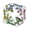
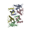
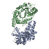
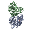
- PDB-6g1n: Crystal structure of the Burkholderia Pseudomallei antitoxin HicB -
+
Open data
ID or keywords:
Loading...
-
Basic information
Entry
Database: PDB / ID: 6g1n
Title
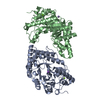
Crystal structure of the Burkholderia Pseudomallei antitoxin HicB
Components
antitoxin HicB
Keywords
ANTITOXIN / The antitoxin HicB which acts as an inhibitor to HicA
Function / homology
HicB-like antitoxin of toxin-antitoxin system / HicB_like antitoxin of bacterial toxin-antitoxin system / TTHA1013/TTHA0281-like / HicB-like antitoxin of toxin-antitoxin system domain-containing protein
Biotechnology and Biological Sciences Research Council
BB/J014400/1
United Kingdom
Citation
Journal: J Biol Chem / Year: 2018 Title: The molecular basis of protein toxin HicA-dependent binding of the protein antitoxin HicB to DNA. Authors: Ashley J Winter / Christopher Williams / Michail N Isupov / Hannah Crocker / Mariya Gromova / Philip Marsh / Oliver J Wilkinson / Mark S Dillingham / Nicholas J Harmer / Richard W Titball / Matthew P Crump / Abstract: Toxin-antitoxin (TA) systems are present in many bacteria and play important roles in bacterial growth, physiology, and pathogenicity. Those that are best studied are the type II TA systems, in which ...Toxin-antitoxin (TA) systems are present in many bacteria and play important roles in bacterial growth, physiology, and pathogenicity. Those that are best studied are the type II TA systems, in which both toxins and antitoxins are proteins. The HicAB system is one of the prototypic TA systems, found in many bacterial species. Complex interactions between the protein toxin (HicA), the protein antitoxin (HicB), and the DNA upstream of the encoding genes regulate the activity of this system, but few structural details are available about how HicA destabilizes the HicB-DNA complex. Here, we determined the X-ray structures of HicB and the HicAB complex to 1.8 and 2.5 Å resolution, respectively, and characterized their DNA interactions. This revealed that HicB forms a tetramer and HicA and HicB form a heterooctameric complex that involves structural reorganization of the C-terminal (DNA-binding) region of HicB. Our observations indicated that HicA has a profound impact on binding of HicB to DNA sequences upstream of in a stoichiometric-dependent way. At low ratios of HicA:HicB, there was no effect on DNA binding, but at higher ratios, the affinity for DNA declined cooperatively, driving dissociation of the HicA:HicB:DNA complex. These results reveal the structural mechanisms by which HicA de-represses the HicB-DNA complex.
Evidence: gel filtration, Analytical SEC showed a tetrameric association, native gel electrophoresis, Native mass spectrometry showed the presence of a tetramer in the gas phase, SAXS, SAXS showed ...Evidence: gel filtration, Analytical SEC showed a tetrameric association, native gel electrophoresis, Native mass spectrometry showed the presence of a tetramer in the gas phase, SAXS, SAXS showed the formation of a tetramer
Type
Name
Symmetry operation
Number
identity operation
1_555
x,y,z
1
Buried area
12550 Å2
ΔGint
-105 kcal/mol
Surface area
26790 Å2
Method
PISA
Unit cell
Length a, b, c (Å)
62.580, 62.580, 173.490
Angle α, β, γ (deg.)
90.00, 90.00, 90.00
Int Tables number
76
Space group name H-M
P41
-
Components
#1: Protein
antitoxinHicB
Mass: 15762.726 Da / Num. of mol.: 4 Source method: isolated from a genetically manipulated source Source: (gene. exp.) Burkholderia pseudomallei (bacteria) / Strain: K96243 / Gene: BPSS0391 / Production host: Escherichia coli (E. coli) / References: UniProt: Q63NA5
 Movie
Movie Controller

 Yorodumi
 Open data
 Basic information
 Components
 Keywords
 Function and homology information
 Burkholderia pseudomallei (bacteria)
 X-RAY DIFFRACTION /
  Authors
 United Kingdom, 1items
  Citation
 Structure visualization
 Downloads & links
 Other downloads


 PDBj
PDBj Assembly
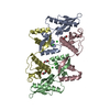

 Mass: 35.453 Da / Num. of mol.: 4 / Source method: obtained synthetically / Formula: Cl
 Mass: 92.094 Da / Num. of mol.: 3 / Source method: obtained synthetically / Formula: C3H8O3
 Mass: 18.015 Da / Num. of mol.: 187 / Source method: isolated from a natural source / Formula: H2O
 Sample preparation
 Processing