 Movie
Movie Controller
[English] 日本語
 Yorodumi
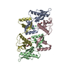
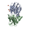
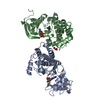
- SASDD45: Type II toxin-antitoxin system HicB family antitoxin - HicB protein -
+ Open data

- Basic information
| Entry | Database: SASBDB / ID: SASDD45 |
|---|---|
 Sample Sample | Type II toxin-antitoxin system HicB family antitoxin - HicB protein
|
| Function / homology | : / HicB-like antitoxin of toxin-antitoxin system / HicB_like antitoxin of bacterial toxin-antitoxin system / TTHA1013/TTHA0281-like / HicB-like antitoxin of toxin-antitoxin system domain-containing protein Function and homology information Function and homology information |
| Biological species |  Burkholderia pseudomallei (bacteria) Burkholderia pseudomallei (bacteria) |
 Citation Citation | Journal: J Biol Chem / Year: 2018 Title: The molecular basis of protein toxin HicA-dependent binding of the protein antitoxin HicB to DNA. Authors: Ashley J Winter / Christopher Williams / Michail N Isupov / Hannah Crocker / Mariya Gromova / Philip Marsh / Oliver J Wilkinson / Mark S Dillingham / Nicholas J Harmer / Richard W Titball / Matthew P Crump /  Abstract: Toxin-antitoxin (TA) systems are present in many bacteria and play important roles in bacterial growth, physiology, and pathogenicity. Those that are best studied are the type II TA systems, in which ...Toxin-antitoxin (TA) systems are present in many bacteria and play important roles in bacterial growth, physiology, and pathogenicity. Those that are best studied are the type II TA systems, in which both toxins and antitoxins are proteins. The HicAB system is one of the prototypic TA systems, found in many bacterial species. Complex interactions between the protein toxin (HicA), the protein antitoxin (HicB), and the DNA upstream of the encoding genes regulate the activity of this system, but few structural details are available about how HicA destabilizes the HicB-DNA complex. Here, we determined the X-ray structures of HicB and the HicAB complex to 1.8 and 2.5 Å resolution, respectively, and characterized their DNA interactions. This revealed that HicB forms a tetramer and HicA and HicB form a heterooctameric complex that involves structural reorganization of the C-terminal (DNA-binding) region of HicB. Our observations indicated that HicA has a profound impact on binding of HicB to DNA sequences upstream of in a stoichiometric-dependent way. At low ratios of HicA:HicB, there was no effect on DNA binding, but at higher ratios, the affinity for DNA declined cooperatively, driving dissociation of the HicA:HicB:DNA complex. These results reveal the structural mechanisms by which HicA de-represses the HicB-DNA complex. |
 Contact author Contact author |
|
- Structure visualization
| Structure viewer | Molecule: MolmilJmol/JSmol |
|---|
- Downloads & links
 SASDD45
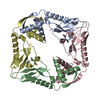


-Models
| Model #1880 |  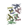 Type: atomic / Chi-square value: 1.97526858112  Search similar-shape structures of this assembly by Omokage search (details) Search similar-shape structures of this assembly by Omokage search (details) |
|---|
-Sample
| Sample | Name: Type II toxin-antitoxin system HicB family antitoxin - HicB protein Specimen concentration: 5 mg/ml |
|---|---|
| Buffer | Name: 25 mM Tris 150 mM NaCl / pH: 7.5 |
| Entity #1010 | Name: HicB / Type: protein Description: Type II toxin-antitoxin system HicB family antitoxin Formula weight: 15.739 / Num. of mol.: 4 / Source: Burkholderia pseudomallei / References: UniProt: Q63NA5 Sequence: MMEFPIAVHK DDGSVYGVTV PDIPGVHSWG ETIDDAIKNT REAIVGHVET LIELGEDVEF TCSTVEELVA KPEYAGAVWA LVSVDLSQLD SKPERINVSI PRFVLHKIDA YVASRHETRS GFLARAALEA LNEGKKHHHH HH |
-Experimental information
| Beam | Instrument name: Diamond Light Source B21 / City: Oxfordshire / 国: UK / Shape: 1 x 5 mm / Type of source: X-ray synchrotron / Wavelength: 0.1 Å / Dist. spec. to detc.: 4.014 mm | |||||||||||||||||||||||||||||||||
|---|---|---|---|---|---|---|---|---|---|---|---|---|---|---|---|---|---|---|---|---|---|---|---|---|---|---|---|---|---|---|---|---|---|---|
| Detector | Name: Pilatus 2M | |||||||||||||||||||||||||||||||||
| Scan |  Measurement date: Jul 28, 2016 / Storage temperature: 4 °C / Cell temperature: 25 °C / Exposure time: 3 sec. / Unit: 1/A /
| |||||||||||||||||||||||||||||||||
| Distance distribution function P(R) | 
| |||||||||||||||||||||||||||||||||
| Result |  Comments: Analysis of the data involved ScÅtter (www.bioisis.net/). The resultant curve was fit with the crystal structure of HicB, PDB Code: 6G1N.
|
