| Entry | Database: PDB / ID: 6g0n
|
|---|
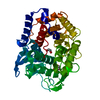
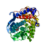
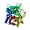
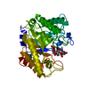
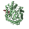
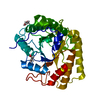
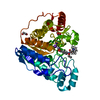
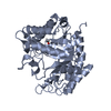
| Title | Crystal Structure of a GH8 catalytic mutant xylohexaose complex xylanase from Teredinibacter turnerae |
|---|
 Components Components | Glycoside hydrolase family 8 domain protein |
|---|
 Keywords Keywords | HYDROLASE / Xylanse / mutant / carbohydrate |
|---|
| Function / homology |  Function and homology information Function and homology information
Glycoside hydrolase, family 8 / Glycosyl hydrolases family 8 / Glycosyltransferase - #10 / Six-hairpin glycosidase-like superfamily / Six-hairpin glycosidase superfamily / Glycosyltransferase / Alpha/alpha barrel / Prokaryotic membrane lipoprotein lipid attachment site profile. / Mainly AlphaSimilarity search - Domain/homology |
|---|
| Biological species |  Teredinibacter turnerae (bacteria) Teredinibacter turnerae (bacteria) |
|---|
| Method |  X-RAY DIFFRACTION / SYNCHROTRON / MOLECULAR REPLACEMENT / Resolution: 1.8 Å X-RAY DIFFRACTION / SYNCHROTRON / MOLECULAR REPLACEMENT / Resolution: 1.8 Å |
|---|
 Authors Authors | Fowler, C.A. / Davies, G.J. / Walton, P.H. |
|---|
| Funding support |  United Kingdom, 1items United Kingdom, 1items | Organization | Grant number | Country |
|---|
| Biotechnology and Biological Sciences Research Council | BB/L001926/1 | United Kingdom |
|
|---|
 Citation Citation | Journal: Acta Crystallogr D Struct Biol / Year: 2018
Title: Structure and function of a glycoside hydrolase family 8 endoxylanase from Teredinibacter turnerae.
Authors: Fowler, C.A. / Hemsworth, G.R. / Cuskin, F. / Hart, S. / Turkenburg, J. / Gilbert, H.J. / Walton, P.H. / Davies, G.J. |
|---|
| History | | Deposition | Mar 19, 2018 | Deposition site: PDBE / Processing site: PDBE |
|---|
| Revision 1.0 | Oct 10, 2018 | Provider: repository / Type: Initial release |
|---|
| Revision 1.1 | Oct 17, 2018 | Group: Data collection / Database references / Category: citation / citation_author
Item: _citation.country / _citation.journal_abbrev ..._citation.country / _citation.journal_abbrev / _citation.journal_id_ASTM / _citation.journal_id_ISSN / _citation.journal_volume / _citation.page_first / _citation.page_last / _citation.pdbx_database_id_PubMed / _citation.title / _citation_author.identifier_ORCID |
|---|
| Revision 2.0 | Jul 29, 2020 | Group: Advisory / Atomic model ...Advisory / Atomic model / Data collection / Derived calculations / Structure summary
Category: atom_site / chem_comp ...atom_site / chem_comp / entity / pdbx_branch_scheme / pdbx_chem_comp_identifier / pdbx_entity_branch / pdbx_entity_branch_descriptor / pdbx_entity_branch_link / pdbx_entity_branch_list / pdbx_entity_nonpoly / pdbx_nonpoly_scheme / pdbx_struct_assembly_gen / pdbx_unobs_or_zero_occ_atoms / pdbx_validate_close_contact / struct_asym / struct_conn / struct_site / struct_site_gen
Item: _atom_site.B_iso_or_equiv / _atom_site.Cartn_x ..._atom_site.B_iso_or_equiv / _atom_site.Cartn_x / _atom_site.Cartn_y / _atom_site.Cartn_z / _atom_site.auth_asym_id / _atom_site.auth_atom_id / _atom_site.auth_seq_id / _atom_site.label_asym_id / _atom_site.label_atom_id / _atom_site.label_entity_id / _atom_site.type_symbol / _chem_comp.name / _chem_comp.type / _pdbx_entity_nonpoly.entity_id / _pdbx_entity_nonpoly.name / _pdbx_struct_assembly_gen.asym_id_list / _pdbx_unobs_or_zero_occ_atoms.auth_atom_id / _pdbx_unobs_or_zero_occ_atoms.label_asym_id / _pdbx_unobs_or_zero_occ_atoms.label_atom_id / _pdbx_validate_close_contact.auth_asym_id_1 / _pdbx_validate_close_contact.auth_atom_id_1 / _pdbx_validate_close_contact.auth_atom_id_2 / _pdbx_validate_close_contact.auth_seq_id_1 / _pdbx_validate_close_contact.auth_seq_id_2 / _struct_conn.pdbx_dist_value / _struct_conn.ptnr1_auth_asym_id / _struct_conn.ptnr1_auth_seq_id / _struct_conn.ptnr1_label_asym_id / _struct_conn.ptnr1_label_atom_id / _struct_conn.ptnr2_auth_asym_id / _struct_conn.ptnr2_auth_seq_id / _struct_conn.ptnr2_label_asym_id / _struct_conn.ptnr2_label_atom_id
Description: Carbohydrate remediation / Provider: repository / Type: Remediation |
|---|
| Revision 2.1 | May 8, 2024 | Group: Data collection / Database references ...Data collection / Database references / Derived calculations / Structure summary
Category: chem_comp / chem_comp_atom ...chem_comp / chem_comp_atom / chem_comp_bond / database_2 / struct_conn
Item: _chem_comp.pdbx_synonyms / _database_2.pdbx_DOI ..._chem_comp.pdbx_synonyms / _database_2.pdbx_DOI / _database_2.pdbx_database_accession / _struct_conn.pdbx_leaving_atom_flag |
|---|
|
|---|
 Movie
Movie Controller

 Yorodumi
 Open data
 Basic information
 Structure visualization
 Downloads & links
 Other downloads
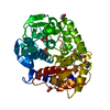


 PDBj
PDBj


 Assembly
 Type: D-saccharide, beta linking / Mass: 150.130 Da / Num. of mol.: 1
 Mass: 92.094 Da / Num. of mol.: 1 / Source method: isolated from a natural source / Formula: C3H8O3
 Mass: 18.015 Da / Num. of mol.: 194 / Source method: isolated from a natural source / Formula: H2O
 Sample preparation
 Processing