 Movie
Movie Controller

[English] 日本語
 Yorodumi
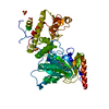
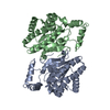
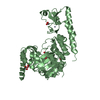
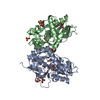
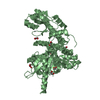
- PDB-6fuk: Crystal structure of UTX complexed with 5-carboxy-8-hydroxyquinoline -
+ Open data

- Basic information
| Entry | Database: PDB / ID: 6fuk | ||||||
|---|---|---|---|---|---|---|---|
| Title | Crystal structure of UTX complexed with 5-carboxy-8-hydroxyquinoline | ||||||
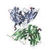
 Components Components | Lysine-specific demethylase 6A | ||||||
 Keywords Keywords | OXIDOREDUCTASE / Jumonji Demethylase / Inhibitor | ||||||
| Function / homology |  Function and homology information Function and homology information[histone H3]-trimethyl-L-lysine27 demethylase / histone H3K27me2/H3K27me3 demethylase activity / Epigenetic regulation of gene expression by MLL3 and MLL4 complexes / MLL3/4 complex / Formation of WDR5-containing histone-modifying complexes / histone methyltransferase complex / histone demethylase activity / Chromatin modifications during the maternal to zygotic transition (MZT) / HDMs demethylate histones / chromatin DNA binding ...[histone H3]-trimethyl-L-lysine27 demethylase / histone H3K27me2/H3K27me3 demethylase activity / Epigenetic regulation of gene expression by MLL3 and MLL4 complexes / MLL3/4 complex / Formation of WDR5-containing histone-modifying complexes / histone methyltransferase complex / histone demethylase activity / Chromatin modifications during the maternal to zygotic transition (MZT) / HDMs demethylate histones / chromatin DNA binding / Activation of anterior HOX genes in hindbrain development during early embryogenesis / heart development / MLL4 and MLL3 complexes regulate expression of PPARG target genes in adipogenesis and hepatic steatosis / regulation of gene expression / RNA polymerase II cis-regulatory region sequence-specific DNA binding / chromatin remodeling / nucleoplasm / metal ion binding / nucleus Similarity search - Function | ||||||
| Biological species |  Homo sapiens (human) Homo sapiens (human) | ||||||
| Method |  X-RAY DIFFRACTION / SYNCHROTRON / MOLECULAR REPLACEMENT / Resolution: 2 Å X-RAY DIFFRACTION / SYNCHROTRON / MOLECULAR REPLACEMENT / Resolution: 2 Å | ||||||
 Authors Authors | Esposito, C. / Sledz, P. / Caflisch, A. | ||||||
 Citation Citation | Journal: J Chem Inf Model / Year: 2018 Title: In Silico Identification of JMJD3 Demethylase Inhibitors. Authors: Esposito, C. / Wiedmer, L. / Caflisch, A. | ||||||
| History |
|
- Structure visualization
| Structure viewer | Molecule: MolmilJmol/JSmol |
|---|
- Downloads & links
-Download
| PDBx/mmCIF format | 6fuk.cif.gz | 121.7 KB | Display | PDBx/mmCIF format |
|---|---|---|---|---|
| PDB format | pdb6fuk.ent.gz | 89.6 KB | Display | PDB format |
| PDBx/mmJSON format | 6fuk.json.gz | Tree view | PDBx/mmJSON format | |
| Others |  Other downloads Other downloads |
-Validation report
| Arichive directory | https://data.pdbj.org/pub/pdb/validation_reports/fu/6fukftp://data.pdbj.org/pub/pdb/validation_reports/fu/6fuk | HTTPS FTP |
|---|
-Related structure data
| Related structure data |  6fulC 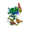 6g8fC  3avsS S: Starting model for refinement C: citing same article ( |
|---|---|
| Similar structure data |


-Links
 PDBj
PDBj


- Assembly
| Deposited unit | 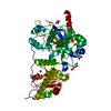
| ||||||||
|---|---|---|---|---|---|---|---|---|---|
| 1 |
| ||||||||
| Unit cell |
|
-Components
-Protein , 1 types, 1 molecules A
| #1: Protein | Mass: 60264.375 Da / Num. of mol.: 1 Source method: isolated from a genetically manipulated source Source: (gene. exp.) Homo sapiens (human) / Gene: KDM6A, UTX / Production host:  References: UniProt: O15550, Oxidoreductases; Acting on paired donors, with incorporation or reduction of molecular oxygen; With 2-oxoglutarate as one donor, and incorporation of one atom of oxygen into each donor |
|---|
-Non-polymers , 7 types, 277 molecules 


| #2: Chemical | ChemComp-MN /  Mass: 54.938 Da / Num. of mol.: 1 / Source method: obtained synthetically / Formula: Mn Mass: 54.938 Da / Num. of mol.: 1 / Source method: obtained synthetically / Formula: Mn | ||||||
|---|---|---|---|---|---|---|---|
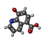
| #3: Chemical | ChemComp-8XQ /  Mass: 189.167 Da / Num. of mol.: 1 / Source method: obtained synthetically / Formula: C10H7NO3 Mass: 189.167 Da / Num. of mol.: 1 / Source method: obtained synthetically / Formula: C10H7NO3 | ||||||
| #4: Chemical | ChemComp-ZN /  Mass: 65.409 Da / Num. of mol.: 1 / Source method: obtained synthetically / Formula: Zn Mass: 65.409 Da / Num. of mol.: 1 / Source method: obtained synthetically / Formula: Zn | ||||||
| #5: Chemical |  Mass: 96.063 Da / Num. of mol.: 2 / Source method: obtained synthetically / Formula: SO4 Mass: 96.063 Da / Num. of mol.: 2 / Source method: obtained synthetically / Formula: SO4#6: Chemical |  Mass: 106.120 Da / Num. of mol.: 2 / Source method: obtained synthetically / Formula: C4H10O3 Mass: 106.120 Da / Num. of mol.: 2 / Source method: obtained synthetically / Formula: C4H10O3#7: Chemical | ChemComp-PG0 / |  Mass: 120.147 Da / Num. of mol.: 1 / Source method: obtained synthetically / Formula: C5H12O3 / Comment: inhibitor, precipitant*YM Mass: 120.147 Da / Num. of mol.: 1 / Source method: obtained synthetically / Formula: C5H12O3 / Comment: inhibitor, precipitant*YM#8: Water | ChemComp-HOH / | Mass: 18.015 Da / Num. of mol.: 269 / Source method: isolated from a natural source / Formula: H2O |
-Experimental details
-Experiment
| Experiment | Method: X-RAY DIFFRACTION / Number of used crystals: 1 |
|---|
- Sample preparation
| Crystal | Density Matthews: 2.58 Å3/Da / Density % sol: 52.33 % |
|---|---|
| Crystal grow | Temperature: 277 K / Method: vapor diffusion, hanging drop / pH: 8.5 Details: 0.1 M Tris-HCl (pH 8.5), Li2 SO4 (0.15-0.25 M), PEG 3350 (20-25% w/v) |
-Data collection
| Diffraction | Mean temperature: 100 K |
|---|---|
| Diffraction source | Source: SYNCHROTRON / Site: SLS  / Beamline: X06SA / Wavelength: 0.999998 Å / Beamline: X06SA / Wavelength: 0.999998 Å |
| Detector | Type: DECTRIS EIGER X 16M / Detector: PIXEL / Date: Nov 6, 2017 |
| Radiation | Protocol: SINGLE WAVELENGTH / Monochromatic (M) / Laue (L): M / Scattering type: x-ray |
| Radiation wavelength | Wavelength: 0.999998 Å / Relative weight: 1 |
| Reflection | Resolution: 2→46.449 Å / Num. obs: 42377 / % possible obs: 98.3 % / Redundancy: 3.7 % / Net I/σ(I): 12.58 |
| Reflection shell | Resolution: 2→5.95 Å / Redundancy: 3.7 % / Mean I/σ(I) obs: 1.67 / Num. unique obs: 6776 / % possible all: 98.4 |
- Processing
| Software |
| ||||||||||||||||||||||||||||||||||||||||||||||||||||||||||||||||||||||||||||||||||||||||||||||||||||||||||||||||
|---|---|---|---|---|---|---|---|---|---|---|---|---|---|---|---|---|---|---|---|---|---|---|---|---|---|---|---|---|---|---|---|---|---|---|---|---|---|---|---|---|---|---|---|---|---|---|---|---|---|---|---|---|---|---|---|---|---|---|---|---|---|---|---|---|---|---|---|---|---|---|---|---|---|---|---|---|---|---|---|---|---|---|---|---|---|---|---|---|---|---|---|---|---|---|---|---|---|---|---|---|---|---|---|---|---|---|---|---|---|---|---|---|---|
| Refinement | Method to determine structure: MOLECULAR REPLACEMENT Starting model: 3AVS Resolution: 2→46.449 Å / SU ML: 0.25 / Cross valid method: FREE R-VALUE / σ(F): 1.35 / Phase error: 26.76
| ||||||||||||||||||||||||||||||||||||||||||||||||||||||||||||||||||||||||||||||||||||||||||||||||||||||||||||||||
| Solvent computation | Shrinkage radii: 0.9 Å / VDW probe radii: 1.11 Å | ||||||||||||||||||||||||||||||||||||||||||||||||||||||||||||||||||||||||||||||||||||||||||||||||||||||||||||||||
| Refinement step | Cycle: LAST / Resolution: 2→46.449 Å
| ||||||||||||||||||||||||||||||||||||||||||||||||||||||||||||||||||||||||||||||||||||||||||||||||||||||||||||||||
| Refine LS restraints |
| ||||||||||||||||||||||||||||||||||||||||||||||||||||||||||||||||||||||||||||||||||||||||||||||||||||||||||||||||
| LS refinement shell |
|
