 Movie
Movie Controller

[English] 日本語
 Yorodumi
- PDB-6fqx: Plasmodium falciparum 6-phosphogluconate dehydrogenase in its apo... -
+ Open data

- Basic information
| Entry | Database: PDB / ID: 6fqx | |||||||||
|---|---|---|---|---|---|---|---|---|---|---|
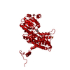
| Title | Plasmodium falciparum 6-phosphogluconate dehydrogenase in its apo form, in complex with its cofactor NADP+ and in complex with its substrate 6-phosphogluconate | |||||||||
 Components Components | 6-phosphogluconate dehydrogenase, decarboxylating | |||||||||
 Keywords Keywords | OXIDOREDUCTASE / Plasmodium / complex / substrate / redoxregulation | |||||||||
| Function / homology |  Function and homology information Function and homology informationphosphogluconate dehydrogenase (NADP+-dependent, decarboxylating) / phosphogluconate dehydrogenase (decarboxylating) activity / D-gluconate metabolic process / pentose-phosphate shunt, oxidative branch / pentose-phosphate shunt / NADP binding / cytosol Similarity search - Function | |||||||||
| Biological species |  | |||||||||
| Method |  X-RAY DIFFRACTION / SYNCHROTRON / MOLECULAR REPLACEMENT / Resolution: 2.8 Å X-RAY DIFFRACTION / SYNCHROTRON / MOLECULAR REPLACEMENT / Resolution: 2.8 Å | |||||||||
 Authors Authors | Fritz-Wolf, K. / Haeussler, K. / Reichmann, M. / Rahlfs, S. / Becker, K. | |||||||||
| Funding support |  Germany, 2items Germany, 2items
| |||||||||
 Citation Citation | Journal: J. Mol. Biol. / Year: 2018 Title: Characterization of Plasmodium falciparum 6-Phosphogluconate Dehydrogenase as an Antimalarial Drug Target. Authors: Haeussler, K. / Fritz-Wolf, K. / Reichmann, M. / Rahlfs, S. / Becker, K. | |||||||||
| History |
|
- Structure visualization
| Structure viewer | Molecule: MolmilJmol/JSmol |
|---|
- Downloads & links
-Download
| PDBx/mmCIF format | 6fqx.cif.gz | 713.3 KB | Display | PDBx/mmCIF format |
|---|---|---|---|---|
| PDB format | pdb6fqx.ent.gz | 596 KB | Display | PDB format |
| PDBx/mmJSON format | 6fqx.json.gz | Tree view | PDBx/mmJSON format | |
| Others |  Other downloads Other downloads |
-Validation report
| Arichive directory | https://data.pdbj.org/pub/pdb/validation_reports/fq/6fqxftp://data.pdbj.org/pub/pdb/validation_reports/fq/6fqx | HTTPS FTP |
|---|
-Related structure data
| Related structure data |  6fqyC  6fqzC  2w90S C: citing same article ( S: Starting model for refinement |
|---|---|
| Similar structure data |

-Links
 PDBj
PDBj


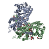
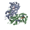
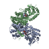
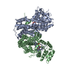
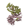
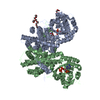
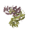
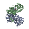
- Assembly
| Deposited unit | 
| ||||||||
|---|---|---|---|---|---|---|---|---|---|
| 1 | 
| ||||||||
| 2 | 
| ||||||||
| 3 | 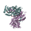
| ||||||||
| 4 | 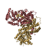
| ||||||||
| Unit cell |
|
-Components
-Protein , 1 types, 8 molecules ABCDEFGH
| #1: Protein | Mass: 53055.875 Da / Num. of mol.: 8 Source method: isolated from a genetically manipulated source Source: (gene. exp.)  References: UniProt: Q8IKT2, phosphogluconate dehydrogenase (NADP+-dependent, decarboxylating) |
|---|
-Non-polymers , 5 types, 124 molecules 


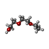

| #2: Chemical | ChemComp-GOL /  Mass: 92.094 Da / Num. of mol.: 14 / Source method: obtained synthetically / Formula: C3H8O3 Mass: 92.094 Da / Num. of mol.: 14 / Source method: obtained synthetically / Formula: C3H8O3#3: Chemical | ChemComp-PEG /  Mass: 106.120 Da / Num. of mol.: 9 / Source method: obtained synthetically / Formula: C4H10O3 Mass: 106.120 Da / Num. of mol.: 9 / Source method: obtained synthetically / Formula: C4H10O3#4: Chemical |  Mass: 150.173 Da / Num. of mol.: 2 / Source method: obtained synthetically / Formula: C6H14O4 Mass: 150.173 Da / Num. of mol.: 2 / Source method: obtained synthetically / Formula: C6H14O4#5: Chemical | ChemComp-AE3 / |  Mass: 134.174 Da / Num. of mol.: 1 / Source method: obtained synthetically / Formula: C6H14O3 Mass: 134.174 Da / Num. of mol.: 1 / Source method: obtained synthetically / Formula: C6H14O3#6: Water | ChemComp-HOH / | Mass: 18.015 Da / Num. of mol.: 98 / Source method: isolated from a natural source / Formula: H2O |
|---|
-Details
| Has protein modification | Y |
|---|
-Experimental details
-Experiment
| Experiment | Method: X-RAY DIFFRACTION / Number of used crystals: 1 |
|---|
- Sample preparation
| Crystal | Density Matthews: 2.4 Å3/Da / Density % sol: 50.72 % |
|---|---|
| Crystal grow | Temperature: 295 K / Method: vapor diffusion, hanging drop / pH: 5.6 Details: 25% PEG 4,000, 15% glycerol, 0.085 M NaCitrat (pH 5.6), and 0.17 M Ammonium acetate |
-Data collection
| Diffraction | Mean temperature: 100 K |
|---|---|
| Diffraction source | Source: SYNCHROTRON / Site: SLS  / Beamline: X10SA / Wavelength: 1 Å / Beamline: X10SA / Wavelength: 1 Å |
| Detector | Type: DECTRIS PILATUS3 S 6M / Detector: PIXEL / Date: Nov 29, 2014 |
| Radiation | Protocol: SINGLE WAVELENGTH / Monochromatic (M) / Laue (L): M / Scattering type: x-ray |
| Radiation wavelength | Wavelength: 1 Å / Relative weight: 1 |
| Reflection | Resolution: 2.8→46.53 Å / Num. obs: 101295 / % possible obs: 99.62 % / Redundancy: 10.3 % / Biso Wilson estimate: 73.5 Å2 / CC1/2: 0.997 / Rmerge(I) obs: 0.1825 / Rpim(I) all: 0.06 / Rrim(I) all: 0.192 / Net I/σ(I): 12.3 |
| Reflection shell | Resolution: 2.8→2.9 Å / Redundancy: 8.6 % / Mean I/σ(I) obs: 0.89 / Num. unique obs: 9878 / CC1/2: 0.25 / % possible all: 97 |
- Processing
| Software |
| |||||||||||||||||||||||||||||||||||||||||||||||||||||||||||||||||||||||||||||||||||||||||||||||||||||||||
|---|---|---|---|---|---|---|---|---|---|---|---|---|---|---|---|---|---|---|---|---|---|---|---|---|---|---|---|---|---|---|---|---|---|---|---|---|---|---|---|---|---|---|---|---|---|---|---|---|---|---|---|---|---|---|---|---|---|---|---|---|---|---|---|---|---|---|---|---|---|---|---|---|---|---|---|---|---|---|---|---|---|---|---|---|---|---|---|---|---|---|---|---|---|---|---|---|---|---|---|---|---|---|---|---|---|---|
| Refinement | Method to determine structure: MOLECULAR REPLACEMENT Starting model: 2w90 Resolution: 2.8→46.528 Å / SU ML: 0.48 / Cross valid method: FREE R-VALUE / σ(F): 1.34 / Phase error: 29.27
| |||||||||||||||||||||||||||||||||||||||||||||||||||||||||||||||||||||||||||||||||||||||||||||||||||||||||
| Solvent computation | Shrinkage radii: 0.9 Å / VDW probe radii: 1.11 Å | |||||||||||||||||||||||||||||||||||||||||||||||||||||||||||||||||||||||||||||||||||||||||||||||||||||||||
| Refinement step | Cycle: LAST / Resolution: 2.8→46.528 Å
| |||||||||||||||||||||||||||||||||||||||||||||||||||||||||||||||||||||||||||||||||||||||||||||||||||||||||
| Refine LS restraints |
| |||||||||||||||||||||||||||||||||||||||||||||||||||||||||||||||||||||||||||||||||||||||||||||||||||||||||
| LS refinement shell |
|
