 Movie
Movie Controller

[English] 日本語
 Yorodumi
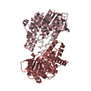
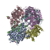
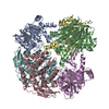
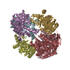
- PDB-6fkx: Crystal structure of an acetyl xylan esterase from a desert metagenome -
+ Open data

- Basic information
| Entry | Database: PDB / ID: 6fkx | ||||||
|---|---|---|---|---|---|---|---|
| Title | Crystal structure of an acetyl xylan esterase from a desert metagenome | ||||||
 Components Components | (Acetyl xylan ...) x 3 | ||||||
 Keywords Keywords | HYDROLASE / Carbohydrate-active enzyme / acetyl xylan esterase / alpha-beta hydrolase / 7-ACA deacetylase | ||||||
| Function / homology | Alpha/Beta hydrolase fold, catalytic domain / Rossmann fold / 3-Layer(aba) Sandwich / Alpha Beta / FORMIC ACID Function and homology information Function and homology information | ||||||
| Biological species | metagenome (others) | ||||||
| Method |  X-RAY DIFFRACTION / SYNCHROTRON / MOLECULAR REPLACEMENT / Resolution: 2.03 Å X-RAY DIFFRACTION / SYNCHROTRON / MOLECULAR REPLACEMENT / Resolution: 2.03 Å | ||||||
 Authors Authors | Adesioye, F.A. / Makhalanyane, T.P. / Vikram, S. / Sewell, B.T. / Schubert, W. / Cowan, D.A. | ||||||
| Funding support |  South Africa, 1items South Africa, 1items
| ||||||
 Citation Citation | Journal: Appl. Environ. Microbiol. / Year: 2018 Title: Structural Characterization and Directed Evolution of a Novel Acetyl Xylan Esterase Reveals Thermostability Determinants of the Carbohydrate Esterase 7 Family. Authors: Adesioye, F.A. / Makhalanyane, T.P. / Vikram, S. / Sewell, B.T. / Schubert, W.D. / Cowan, D.A. | ||||||
| History |
|
- Structure visualization
| Structure viewer | Molecule: MolmilJmol/JSmol |
|---|
- Downloads & links
-Download
| PDBx/mmCIF format | 6fkx.cif.gz | 795.4 KB | Display | PDBx/mmCIF format |
|---|---|---|---|---|
| PDB format | pdb6fkx.ent.gz | 646.7 KB | Display | PDB format |
| PDBx/mmJSON format | 6fkx.json.gz | Tree view | PDBx/mmJSON format | |
| Others |  Other downloads Other downloads |
-Validation report
| Arichive directory | https://data.pdbj.org/pub/pdb/validation_reports/fk/6fkxftp://data.pdbj.org/pub/pdb/validation_reports/fk/6fkx | HTTPS FTP |
|---|
-Related structure data
| Related structure data |  3fcyS S: Starting model for refinement |
|---|---|
| Similar structure data |
-Links
 PDBj
PDBj- Assembly
| Deposited unit | 
| ||||||||||||
|---|---|---|---|---|---|---|---|---|---|---|---|---|---|
| 1 |
| ||||||||||||
| Unit cell |
|
-Components
-Acetyl xylan ... , 3 types, 6 molecules ABDEFC
| #1: Protein | Mass: 36036.684 Da / Num. of mol.: 1 Source method: isolated from a genetically manipulated source Details: Namib Desert Soil Hypolith / Source: (gene. exp.) metagenome (others) / Plasmid: pET28a / Details (production host): Circular with 6X His-tags / Production host:  | ||
|---|---|---|---|
| #2: Protein | Mass: 35760.395 Da / Num. of mol.: 4 Source method: isolated from a genetically manipulated source Details: Namib Desert Soil Hypolith / Source: (gene. exp.) metagenome (others) / Plasmid: pET28a / Details (production host): Circular 6X His-tag / Production host: #3: Protein | | Mass: 35426.008 Da / Num. of mol.: 1 Source method: isolated from a genetically manipulated source Details: Namib Desert Soil Hypolith / Source: (gene. exp.) metagenome (others) / Plasmid: pET28a / Details (production host): Circular, 6X His-tag / Production host: |
-Non-polymers , 5 types, 2285 molecules 


| #4: Chemical | ChemComp-NA /  Mass: 22.990 Da / Num. of mol.: 6 / Source method: obtained synthetically / Formula: Na Mass: 22.990 Da / Num. of mol.: 6 / Source method: obtained synthetically / Formula: Na#5: Chemical | ChemComp-MES /  Mass: 195.237 Da / Num. of mol.: 13 / Source method: obtained synthetically / Formula: C6H13NO4S / Comment: pH buffer*YM Mass: 195.237 Da / Num. of mol.: 13 / Source method: obtained synthetically / Formula: C6H13NO4S / Comment: pH buffer*YM#6: Chemical | ChemComp-SO4 / |  Mass: 96.063 Da / Num. of mol.: 1 / Source method: obtained synthetically / Formula: SO4 Mass: 96.063 Da / Num. of mol.: 1 / Source method: obtained synthetically / Formula: SO4#7: Chemical | ChemComp-FMT / |  Mass: 46.025 Da / Num. of mol.: 1 / Source method: obtained synthetically / Formula: CH2O2 Mass: 46.025 Da / Num. of mol.: 1 / Source method: obtained synthetically / Formula: CH2O2#8: Water | ChemComp-HOH / | Mass: 18.015 Da / Num. of mol.: 2264 / Source method: isolated from a natural source / Formula: H2O |
|---|
-Experimental details
-Experiment
| Experiment | Method: X-RAY DIFFRACTION / Number of used crystals: 1 |
|---|
- Sample preparation
| Crystal | Density Matthews: 2.34 Å3/Da / Density % sol: 47.38 % |
|---|---|
| Crystal grow | Temperature: 291.15 K / Method: vapor diffusion, sitting drop / pH: 8.5 Details: 0.1 M 2-(N-morpholino) ethanesulfonic acid (MES) buffers pH 8.5 and 25% PEG 8000 (w/v) PH range: 6.5-8.5 |
-Data collection
| Diffraction | Mean temperature: 100 K |
|---|---|
| Diffraction source | Source: SYNCHROTRON / Site: ESRF  / Beamline: ID23-1 / Wavelength: 0.972 Å / Beamline: ID23-1 / Wavelength: 0.972 Å |
| Detector | Type: DECTRIS PILATUS 6M / Detector: PIXEL / Date: Jul 9, 2016 |
| Radiation | Monochromator: Si 111 / Protocol: SINGLE WAVELENGTH / Monochromatic (M) / Laue (L): M / Scattering type: x-ray |
| Radiation wavelength | Wavelength: 0.972 Å / Relative weight: 1 |
| Reflection | Resolution: 2.026→89.23 Å / Num. obs: 130734 / % possible obs: 99.95 % / Redundancy: 2 % / Rmerge(I) obs: 0.06564 / Rpim(I) all: 0.06564 / Rrim(I) all: 0.09283 / Net I/σ(I): 10.02 |
| Reflection shell | Resolution: 2.026→2.099 Å / Rmerge(I) obs: 0.3479 / Rpim(I) all: 0.3479 / Rrim(I) all: 0.4921 |
- Processing
| Software |
| ||||||||||||||||
|---|---|---|---|---|---|---|---|---|---|---|---|---|---|---|---|---|---|
| Refinement | Method to determine structure: MOLECULAR REPLACEMENT Starting model: 3FCY Resolution: 2.03→89.23 Å / Cross valid method: FREE R-VALUE
| ||||||||||||||||
| Refinement step | Cycle: LAST / Resolution: 2.03→89.23 Å
|
