 Movie
Movie Controller

[English] 日本語
 Yorodumi
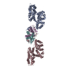
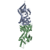
- PDB-6fj3: High resolution crystal structure of parathyroid hormone 1 recept... -
+ Open data

- Basic information
| Entry | Database: PDB / ID: 6fj3 | |||||||||
|---|---|---|---|---|---|---|---|---|---|---|
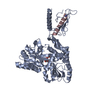
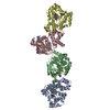
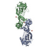
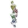
| Title | High resolution crystal structure of parathyroid hormone 1 receptor in complex with a peptide agonist. | |||||||||
 Components Components |
| |||||||||
 Keywords Keywords | MEMBRANE PROTEIN / GPCR / cell signalling / 7TM | |||||||||
| Function / homology |  Function and homology information Function and homology informationparathyroid hormone receptor binding / type 1 parathyroid hormone receptor binding / negative regulation of apoptotic process in bone marrow cell / positive regulation of osteoclast proliferation / positive regulation of cell proliferation in bone marrow / parathyroid hormone receptor activity / alpha-1,4-glucan glucosyltransferase (UDP-glucose donor) activity / positive regulation of signal transduction / magnesium ion homeostasis / phosphate ion homeostasis ...parathyroid hormone receptor binding / type 1 parathyroid hormone receptor binding / negative regulation of apoptotic process in bone marrow cell / positive regulation of osteoclast proliferation / positive regulation of cell proliferation in bone marrow / parathyroid hormone receptor activity / alpha-1,4-glucan glucosyltransferase (UDP-glucose donor) activity / positive regulation of signal transduction / magnesium ion homeostasis / phosphate ion homeostasis / G protein-coupled peptide receptor activity / Class B/2 (Secretin family receptors) / osteoblast development / peptide hormone receptor binding / bone mineralization / positive regulation of inositol phosphate biosynthetic process / peptide hormone binding / positive regulation of glycogen biosynthetic process / chondrocyte differentiation / bone resorption / positive regulation of bone mineralization / G protein-coupled receptor signaling pathway, coupled to cyclic nucleotide second messenger / cell maturation / homeostasis of number of cells within a tissue / positive regulation of D-glucose import across plasma membrane / skeletal system development / hormone activity / G protein-coupled receptor activity / intracellular calcium ion homeostasis / adenylate cyclase-modulating G protein-coupled receptor signaling pathway / cell-cell signaling / adenylate cyclase-activating G protein-coupled receptor signaling pathway / transcription by RNA polymerase II / G alpha (s) signalling events / basolateral plasma membrane / phospholipase C-activating G protein-coupled receptor signaling pathway / in utero embryonic development / cell surface receptor signaling pathway / signaling receptor complex / cell population proliferation / apical plasma membrane / G protein-coupled receptor signaling pathway / negative regulation of cell population proliferation / negative regulation of gene expression / positive regulation of cell population proliferation / nucleolus / protein homodimerization activity / positive regulation of transcription by RNA polymerase II / : / nucleus / plasma membrane / cytoplasm / cytosol Similarity search - Function | |||||||||
| Biological species |  Homo sapiens (human) Homo sapiens (human)  Pyrococcus abyssi (archaea) Pyrococcus abyssi (archaea) | |||||||||
| Method |  X-RAY DIFFRACTION / SYNCHROTRON / MOLECULAR REPLACEMENT / Resolution: 2.5 Å X-RAY DIFFRACTION / SYNCHROTRON / MOLECULAR REPLACEMENT / Resolution: 2.5 Å | |||||||||
 Authors Authors | Ehrenmann, J. / Schoppe, J. / Klenk, C. / Rappas, M. / Kummer, L. / Dore, A.S. / Pluckthun, A. | |||||||||
 Citation Citation | Journal: Nat. Struct. Mol. Biol. / Year: 2018 Title: High-resolution crystal structure of parathyroid hormone 1 receptor in complex with a peptide agonist. Authors: Ehrenmann, J. / Schoppe, J. / Klenk, C. / Rappas, M. / Kummer, L. / Dore, A.S. / Pluckthun, A. | |||||||||
| History |
|
- Structure visualization
| Structure viewer | Molecule: MolmilJmol/JSmol |
|---|
- Downloads & links
-Download
| PDBx/mmCIF format | 6fj3.cif.gz | 281.4 KB | Display | PDBx/mmCIF format |
|---|---|---|---|---|
| PDB format | pdb6fj3.ent.gz | 223.9 KB | Display | PDB format |
| PDBx/mmJSON format | 6fj3.json.gz | Tree view | PDBx/mmJSON format | |
| Others |  Other downloads Other downloads |
-Validation report
| Arichive directory | https://data.pdbj.org/pub/pdb/validation_reports/fj/6fj3ftp://data.pdbj.org/pub/pdb/validation_reports/fj/6fj3 | HTTPS FTP |
|---|
-Related structure data


-Links
 PDBj
PDBj


- Assembly
| Deposited unit | 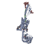
| ||||||||
|---|---|---|---|---|---|---|---|---|---|
| 1 |
| ||||||||
| Unit cell |
|
-Components
-Protein / Protein/peptide , 2 types, 2 molecules AB
| #1: Protein | Mass: 68389.203 Da / Num. of mol.: 1 Mutation: Y191C, K240M, L300A, M312K, V334I, K359N, L407A, A426L, Q440R, I458A,Y191C, K240M, L300A, M312K, V334I, K359N, L407A, A426L, Q440R, I458A,Y191C, K240M, L300A, M312K, V334I, K359N, L407A, ...Mutation: Y191C, K240M, L300A, M312K, V334I, K359N, L407A, A426L, Q440R, I458A,Y191C, K240M, L300A, M312K, V334I, K359N, L407A, A426L, Q440R, I458A,Y191C, K240M, L300A, M312K, V334I, K359N, L407A, A426L, Q440R, I458A,Y191C, K240M, L300A, M312K, V334I, K359N, L407A, A426L, Q440R, I458A Source method: isolated from a genetically manipulated source Source: (gene. exp.) Homo sapiens (human), (gene. exp.) Pyrococcus abyssi (strain GE5 / Orsay) (archaea)Gene: PTH1R, PTHR, PTHR1, PAB2292 / Strain: GE5 / Orsay / Production host:   Spodoptera frugiperda (fall armyworm) / References: UniProt: Q03431, UniProt: Q9V2J8 Spodoptera frugiperda (fall armyworm) / References: UniProt: Q03431, UniProt: Q9V2J8 |
|---|---|
| #2: Protein/peptide | Mass: 4277.009 Da / Num. of mol.: 1 / Mutation: N41Q, G43A, H45W Source method: isolated from a genetically manipulated source Source: (gene. exp.) Spodoptera frugiperda (fall armyworm) / References: UniProt: Q27IM2 |
-Sugars , 4 types, 6 molecules 
| #3: Polysaccharide | alpha-D-mannopyranose-(1-3)-[alpha-D-mannopyranose-(1-6)]beta-D-mannopyranose-(1-4)-2-acetamido-2- ...alpha-D-mannopyranose-(1-3)-[alpha-D-mannopyranose-(1-6)]beta-D-mannopyranose-(1-4)-2-acetamido-2-deoxy-beta-D-glucopyranose-(1-4)-[alpha-L-fucopyranose-(1-6)]2-acetamido-2-deoxy-beta-D-glucopyranose Source method: isolated from a genetically manipulated source |
|---|---|
| #4: Polysaccharide | beta-D-mannopyranose-(1-4)-2-acetamido-2-deoxy-beta-D-glucopyranose-(1-4)-[alpha-L-fucopyranose-(1- ...beta-D-mannopyranose-(1-4)-2-acetamido-2-deoxy-beta-D-glucopyranose-(1-4)-[alpha-L-fucopyranose-(1-6)]2-acetamido-2-deoxy-beta-D-glucopyranose Source method: isolated from a genetically manipulated source |
| #5: Polysaccharide | alpha-D-mannopyranose-(1-6)-beta-D-mannopyranose-(1-4)-2-acetamido-2-deoxy-beta-D-glucopyranose-(1- ...alpha-D-mannopyranose-(1-6)-beta-D-mannopyranose-(1-4)-2-acetamido-2-deoxy-beta-D-glucopyranose-(1-4)-2-acetamido-2-deoxy-beta-D-glucopyranose Source method: isolated from a genetically manipulated source |
| #6: Sugar |  Type: D-saccharide, alpha linking / Mass: 180.156 Da / Num. of mol.: 3 Type: D-saccharide, alpha linking / Mass: 180.156 Da / Num. of mol.: 3Source method: isolated from a genetically manipulated source Formula: C6H12O6 |
-Non-polymers , 5 types, 168 molecules 


| #7: Chemical | ChemComp-OLA /  Mass: 282.461 Da / Num. of mol.: 9 Mass: 282.461 Da / Num. of mol.: 9Source method: isolated from a genetically manipulated source Formula: C18H34O2 #8: Chemical | ChemComp-ACY / |  Mass: 60.052 Da / Num. of mol.: 1 Mass: 60.052 Da / Num. of mol.: 1Source method: isolated from a genetically manipulated source Formula: C2H4O2 #9: Chemical | ChemComp-PG4 / |  Mass: 194.226 Da / Num. of mol.: 1 / Source method: obtained synthetically / Formula: C8H18O5 / Comment: precipitant*YM Mass: 194.226 Da / Num. of mol.: 1 / Source method: obtained synthetically / Formula: C8H18O5 / Comment: precipitant*YM#10: Chemical | ChemComp-CL / |  Mass: 35.453 Da / Num. of mol.: 1 / Source method: obtained synthetically / Formula: Cl Mass: 35.453 Da / Num. of mol.: 1 / Source method: obtained synthetically / Formula: Cl#11: Water | ChemComp-HOH / | Mass: 18.015 Da / Num. of mol.: 156 / Source method: isolated from a natural source / Formula: H2O |
|---|
-Experimental details
-Experiment
| Experiment | Method: X-RAY DIFFRACTION / Number of used crystals: 1 |
|---|
- Sample preparation
| Crystal | Density Matthews: 3.47 Å3/Da / Density % sol: 64.53 % |
|---|---|
| Crystal grow | Temperature: 293 K / Method: lipidic cubic phase / pH: 6 Details: 0.1 M sodium citrate pH 6.0, 0.3 M sodium acetate, 31% PEG400, 20 uM E-PTH(1-34) |
-Data collection
| Diffraction | Mean temperature: 100 K |
|---|---|
| Diffraction source | Source: SYNCHROTRON / Site: SLS  / Beamline: X06SA / Wavelength: 1 Å / Beamline: X06SA / Wavelength: 1 Å |
| Detector | Type: DECTRIS EIGER X 16M / Detector: PIXEL / Date: Sep 4, 2017 |
| Radiation | Monochromator: LN2 cooled fixed exit Si(111) monochromator / Protocol: SINGLE WAVELENGTH / Monochromatic (M) / Laue (L): M / Scattering type: x-ray |
| Radiation wavelength | Wavelength: 1 Å / Relative weight: 1 |
| Reflection | Resolution: 2.5→49.39 Å / Num. obs: 33759 / % possible obs: 99.8 % / Redundancy: 5.3 % / Biso Wilson estimate: 50.93 Å2 / CC1/2: 0.994 / Rmerge(I) obs: 0.153 / Rpim(I) all: 0.103 / Net I/σ(I): 5.2 |
| Reflection shell | Resolution: 2.5→2.6 Å / Redundancy: 4.7 % / Rmerge(I) obs: 1.662 / Mean I/σ(I) obs: 0.9 / Num. unique obs: 3825 / CC1/2: 0.501 / Rpim(I) all: 1.214 / % possible all: 99.3 |
- Processing
| Software |
| |||||||||||||||||||||||||||||||||||||||||||||||||||||||||||||||||||||||||||||||||||||||||||||||||||||||||||||||||||||||||||||
|---|---|---|---|---|---|---|---|---|---|---|---|---|---|---|---|---|---|---|---|---|---|---|---|---|---|---|---|---|---|---|---|---|---|---|---|---|---|---|---|---|---|---|---|---|---|---|---|---|---|---|---|---|---|---|---|---|---|---|---|---|---|---|---|---|---|---|---|---|---|---|---|---|---|---|---|---|---|---|---|---|---|---|---|---|---|---|---|---|---|---|---|---|---|---|---|---|---|---|---|---|---|---|---|---|---|---|---|---|---|---|---|---|---|---|---|---|---|---|---|---|---|---|---|---|---|---|
| Refinement | Method to determine structure: MOLECULAR REPLACEMENT Starting model: 5EE7, 3C4M Resolution: 2.5→49.386 Å / SU ML: 0.38 / Cross valid method: FREE R-VALUE / σ(F): 1.97 / Phase error: 28.73 / Stereochemistry target values: ML
| |||||||||||||||||||||||||||||||||||||||||||||||||||||||||||||||||||||||||||||||||||||||||||||||||||||||||||||||||||||||||||||
| Solvent computation | Shrinkage radii: 0.9 Å / VDW probe radii: 1.11 Å / Solvent model: FLAT BULK SOLVENT MODEL | |||||||||||||||||||||||||||||||||||||||||||||||||||||||||||||||||||||||||||||||||||||||||||||||||||||||||||||||||||||||||||||
| Refinement step | Cycle: LAST / Resolution: 2.5→49.386 Å
| |||||||||||||||||||||||||||||||||||||||||||||||||||||||||||||||||||||||||||||||||||||||||||||||||||||||||||||||||||||||||||||
| Refine LS restraints |
| |||||||||||||||||||||||||||||||||||||||||||||||||||||||||||||||||||||||||||||||||||||||||||||||||||||||||||||||||||||||||||||
| LS refinement shell |
| |||||||||||||||||||||||||||||||||||||||||||||||||||||||||||||||||||||||||||||||||||||||||||||||||||||||||||||||||||||||||||||
| Refinement TLS params. | Method: refined / Refine-ID: X-RAY DIFFRACTION
| |||||||||||||||||||||||||||||||||||||||||||||||||||||||||||||||||||||||||||||||||||||||||||||||||||||||||||||||||||||||||||||
| Refinement TLS group |
|
