 Movie
Movie Controller

[English] 日本語
 Yorodumi
- PDB-6fgk: Crystal structure of the small alarmone synthethase 2 from Bacill... -
+ Open data

- Basic information
| Entry | Database: PDB / ID: 6fgk | ||||||
|---|---|---|---|---|---|---|---|
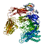
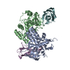
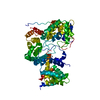
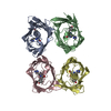
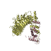
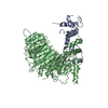
| Title | Crystal structure of the small alarmone synthethase 2 from Bacillus subtilis | ||||||
 Components Components | GTP pyrophosphokinase YwaC | ||||||
 Keywords Keywords | TRANSFERASE / Stringent response / alarmone / SAS2 / RelP | ||||||
| Function / homology |  Function and homology information Function and homology informationGTP diphosphokinase / GTP diphosphokinase activity / guanosine tetraphosphate biosynthetic process / kinase activity / GTP binding / ATP binding Similarity search - Function | ||||||
| Biological species |  | ||||||
| Method |  X-RAY DIFFRACTION / SYNCHROTRON / MOLECULAR REPLACEMENT / Resolution: 3.2 Å X-RAY DIFFRACTION / SYNCHROTRON / MOLECULAR REPLACEMENT / Resolution: 3.2 Å | ||||||
 Authors Authors | Bange, G. / Vogt, M. / Steinchen, W. / Altegoer, F. | ||||||
 Citation Citation | Journal: Sci Rep / Year: 2018 Title: Structural and mechanistic divergence of the small (p)ppGpp synthetases RelP and RelQ. Authors: Steinchen, W. / Vogt, M.S. / Altegoer, F. / Giammarinaro, P.I. / Horvatek, P. / Wolz, C. / Bange, G. | ||||||
| History |
|
- Structure visualization
| Structure viewer | Molecule: MolmilJmol/JSmol |
|---|
- Downloads & links
-Download
| PDBx/mmCIF format | 6fgk.cif.gz | 157.9 KB | Display | PDBx/mmCIF format |
|---|---|---|---|---|
| PDB format | pdb6fgk.ent.gz | 126.7 KB | Display | PDB format |
| PDBx/mmJSON format | 6fgk.json.gz | Tree view | PDBx/mmJSON format | |
| Others |  Other downloads Other downloads |
-Validation report
| Arichive directory | https://data.pdbj.org/pub/pdb/validation_reports/fg/6fgkftp://data.pdbj.org/pub/pdb/validation_reports/fg/6fgk | HTTPS FTP |
|---|
-Related structure data
| Related structure data |  6fgjC 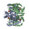 6fgxC 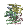 5decS S: Starting model for refinement C: citing same article ( |
|---|---|
| Similar structure data |


-Links
 PDBj
PDBj- Assembly
| Deposited unit | 
| ||||||||
|---|---|---|---|---|---|---|---|---|---|
| 1 |
| ||||||||
| Unit cell |
|
-Components
| #1: Protein | Mass: 24638.586 Da / Num. of mol.: 4 Source method: isolated from a genetically manipulated source Source: (gene. exp.) Strain: 168 / Gene: ywaC, BSU38480, ipa-7d / Production host: |
|---|
-Experimental details
-Experiment
| Experiment | Method: X-RAY DIFFRACTION / Number of used crystals: 1 |
|---|
- Sample preparation
| Crystal | Density Matthews: 2.61 Å3/Da / Density % sol: 52.8 % |
|---|---|
| Crystal grow | Temperature: 294 K / Method: vapor diffusion, sitting drop / Details: 0.1 M CHES pH 9.5, 30% (w/v) PEG 3000 |
-Data collection
| Diffraction | Mean temperature: 100 K |
|---|---|
| Diffraction source | Source: SYNCHROTRON / Site: ESRF  / Beamline: ID23-1 / Wavelength: 0.987 Å / Beamline: ID23-1 / Wavelength: 0.987 Å |
| Detector | Type: DECTRIS PILATUS 6M / Detector: PIXEL / Date: Oct 2, 2016 |
| Radiation | Protocol: SINGLE WAVELENGTH / Monochromatic (M) / Laue (L): M / Scattering type: x-ray |
| Radiation wavelength | Wavelength: 0.987 Å / Relative weight: 1 |
| Reflection | Resolution: 3.2→47.28 Å / Num. obs: 16011 / % possible obs: 92.2 % / Redundancy: 4.1 % / CC1/2: 0.98 / Rmerge(I) obs: 0.169 / Net I/σ(I): 3.7 |
| Reflection shell | Resolution: 3.2→3.42 Å / Redundancy: 4.2 % / Rmerge(I) obs: 0.572 / Mean I/σ(I) obs: 0.9 / Num. unique obs: 1561 / CC1/2: 0.72 / % possible all: 92.7 |
- Processing
| Software |
| |||||||||||||||||||||||||||||||||||||||||||||||||
|---|---|---|---|---|---|---|---|---|---|---|---|---|---|---|---|---|---|---|---|---|---|---|---|---|---|---|---|---|---|---|---|---|---|---|---|---|---|---|---|---|---|---|---|---|---|---|---|---|---|---|
| Refinement | Method to determine structure: MOLECULAR REPLACEMENT Starting model: 5DEC Resolution: 3.2→46.281 Å / SU ML: 0.47 / Cross valid method: FREE R-VALUE / σ(F): 1.36 / Phase error: 31.38
| |||||||||||||||||||||||||||||||||||||||||||||||||
| Solvent computation | Shrinkage radii: 0.9 Å / VDW probe radii: 1.11 Å | |||||||||||||||||||||||||||||||||||||||||||||||||
| Refinement step | Cycle: LAST / Resolution: 3.2→46.281 Å
| |||||||||||||||||||||||||||||||||||||||||||||||||
| Refine LS restraints |
| |||||||||||||||||||||||||||||||||||||||||||||||||
| LS refinement shell |
|
