 Movie
Movie Controller


+ Open data

- Basic information
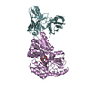
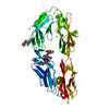
| Entry | Database: PDB / ID: 6ffh | ||||||
|---|---|---|---|---|---|---|---|
| Title | Crystal Structure of mGluR5 in complex with Fenobam at 2.65 A | ||||||
 Components Components | Metabotropic glutamate receptor 5,Endolysin | ||||||
 Keywords Keywords | MEMBRANE PROTEIN / 7TM / RECEPTOR / GPCR / MEMBRANE-PROTEIN / SIGNALING PROTEIN | ||||||
| Function / homology |  Function and homology information Function and homology informationA2A adenosine receptor binding / sensory perception of hot stimulus / negative regulation of dendritic spine morphogenesis / : / G protein-coupled receptor activity involved in regulation of postsynaptic membrane potential / adenylate cyclase inhibiting G protein-coupled glutamate receptor activity / protein localization to nuclear inner membrane / positive regulation of cellular response to hypoxia / phospholipase C-activating G protein-coupled glutamate receptor signaling pathway / operant conditioning ...A2A adenosine receptor binding / sensory perception of hot stimulus / negative regulation of dendritic spine morphogenesis / : / G protein-coupled receptor activity involved in regulation of postsynaptic membrane potential / adenylate cyclase inhibiting G protein-coupled glutamate receptor activity / protein localization to nuclear inner membrane / positive regulation of cellular response to hypoxia / phospholipase C-activating G protein-coupled glutamate receptor signaling pathway / operant conditioning / positive regulation of long-term neuronal synaptic plasticity / positive regulation of sensory perception of pain / negative regulation of excitatory postsynaptic potential / positive regulation of dopamine secretion / desensitization of G protein-coupled receptor signaling pathway / G protein-coupled glutamate receptor signaling pathway / positive regulation of neural precursor cell proliferation / Class C/3 (Metabotropic glutamate/pheromone receptors) / nuclear inner membrane / glutamate receptor activity / Neurexins and neuroligins / astrocyte projection / response to corticosterone / response to morphine / temperature homeostasis / protein tyrosine kinase activator activity / viral release from host cell by cytolysis / conditioned place preference / regulation of synaptic transmission, glutamatergic / peptidoglycan catabolic process / regulation of long-term synaptic depression / positive regulation of calcium-mediated signaling / protein tyrosine kinase binding / dendritic shaft / response to amphetamine / locomotory behavior / synapse organization / postsynaptic density membrane / Schaffer collateral - CA1 synapse / cognition / G protein-coupled receptor activity / cellular response to amyloid-beta / cell wall macromolecule catabolic process / lysozyme / lysozyme activity / positive regulation of cytosolic calcium ion concentration / dendritic spine / G alpha (q) signalling events / host cell cytoplasm / chemical synaptic transmission / response to ethanol / learning or memory / positive regulation of MAPK cascade / defense response to bacterium / response to antibiotic / neuronal cell body / regulation of DNA-templated transcription / dendrite / glutamatergic synapse / identical protein binding / plasma membrane / cytoplasm Similarity search - Function | ||||||
| Biological species |  Homo sapiens (human) Homo sapiens (human) Enterobacteria phage T4 (virus) Enterobacteria phage T4 (virus) | ||||||
| Method |  X-RAY DIFFRACTION / SYNCHROTRON / MOLECULAR REPLACEMENT / molecular replacement / Resolution: 2.65 Å X-RAY DIFFRACTION / SYNCHROTRON / MOLECULAR REPLACEMENT / molecular replacement / Resolution: 2.65 Å | ||||||
 Authors Authors | Christopher, J.A. / Orgovan, Z. / Congreve, M. / Dore, A.S. / Errey, J.C. / Marshall, F.H. / Mason, J.S. / Okrasa, K. / Rucktooa, P. / Serrano-Vega, M.J. ...Christopher, J.A. / Orgovan, Z. / Congreve, M. / Dore, A.S. / Errey, J.C. / Marshall, F.H. / Mason, J.S. / Okrasa, K. / Rucktooa, P. / Serrano-Vega, M.J. / Ferenczy, G.G. / Keseru, G.M. | ||||||
 Citation Citation | Journal: J.Med.Chem. / Year: 2019 Title: Structure-Based Optimization Strategies for G Protein-Coupled Receptor (GPCR) Allosteric Modulators: A Case Study from Analyses of New Metabotropic Glutamate Receptor 5 (mGlu5) X-ray Structures. Authors: Christopher, J.A. / Orgovan, Z. / Congreve, M. / Dore, A.S. / Errey, J.C. / Marshall, F.H. / Mason, J.S. / Okrasa, K. / Rucktooa, P. / Serrano-Vega, M.J. / Ferenczy, G.G. / Keseru, G.M. | ||||||
| History |
|
- Structure visualization
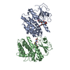
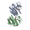
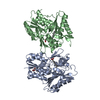
| Structure viewer | Molecule: MolmilJmol/JSmol |
|---|
- Downloads & links
-Download
| PDBx/mmCIF format | 6ffh.cif.gz | 184.7 KB | Display | PDBx/mmCIF format |
|---|---|---|---|---|
| PDB format | pdb6ffh.ent.gz | 144.2 KB | Display | PDB format |
| PDBx/mmJSON format | 6ffh.json.gz | Tree view | PDBx/mmJSON format | |
| Others |  Other downloads Other downloads |
-Validation report
| Arichive directory | https://data.pdbj.org/pub/pdb/validation_reports/ff/6ffhftp://data.pdbj.org/pub/pdb/validation_reports/ff/6ffh | HTTPS FTP |
|---|
-Related structure data
| Related structure data |  6ffiC  4oo9S S: Starting model for refinement C: citing same article ( |
|---|---|
| Similar structure data |


-Links
 PDBj
PDBj


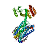
- Assembly
| Deposited unit | 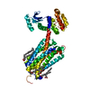
| ||||||||
|---|---|---|---|---|---|---|---|---|---|
| 1 |
| ||||||||
| Unit cell |
|
-Components
-Protein , 1 types, 1 molecules A
| #1: Protein | Mass: 49859.586 Da / Num. of mol.: 1 / Fragment: MGLUR5 / Mutation: C54T C97A E579A N667Y I669A G675M T742A S753A Source method: isolated from a genetically manipulated source Details: Chimeric construct of Human mGLU5 (GRM5) with a Bacteriophage T4 Lysozyme (P00720) insertion in intracellular loop 2 between residues LYS678 and LYS679. Source: (gene. exp.) Homo sapiens (human), (gene. exp.) Enterobacteria phage T4 (virus)Gene: GRM5, GPRC1E, MGLUR5 / Cell line (production host): Sf21 / Production host:   Spodoptera frugiperda (fall armyworm) / References: UniProt: P41594, UniProt: P00720, lysozyme Spodoptera frugiperda (fall armyworm) / References: UniProt: P41594, UniProt: P00720, lysozyme |
|---|
-Non-polymers , 5 types, 21 molecules 

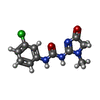


| #2: Chemical | ChemComp-OLA /  Mass: 282.461 Da / Num. of mol.: 5 / Fragment: T4L / Mutation: C54T C97A Mass: 282.461 Da / Num. of mol.: 5 / Fragment: T4L / Mutation: C54T C97ASource method: isolated from a genetically manipulated source Formula: C18H34O2 / References: lysozyme #3: Chemical | ChemComp-MES / |  Mass: 195.237 Da / Num. of mol.: 1 / Source method: obtained synthetically / Formula: C6H13NO4S / Comment: pH buffer*YM Mass: 195.237 Da / Num. of mol.: 1 / Source method: obtained synthetically / Formula: C6H13NO4S / Comment: pH buffer*YM#4: Chemical | ChemComp-D7W / |  Mass: 266.684 Da / Num. of mol.: 1 / Source method: obtained synthetically / Formula: C11H11ClN4O2 Mass: 266.684 Da / Num. of mol.: 1 / Source method: obtained synthetically / Formula: C11H11ClN4O2#5: Chemical | ChemComp-OLC / ( |  Mass: 356.540 Da / Num. of mol.: 1 / Source method: obtained synthetically / Formula: C21H40O4 Mass: 356.540 Da / Num. of mol.: 1 / Source method: obtained synthetically / Formula: C21H40O4#6: Water | ChemComp-HOH / | Mass: 18.015 Da / Num. of mol.: 13 / Source method: isolated from a natural source / Formula: H2O |
|---|
-Experimental details
-Experiment
| Experiment | Method: X-RAY DIFFRACTION / Number of used crystals: 1 |
|---|
- Sample preparation
| Crystal | Density Matthews: 2.68 Å3/Da / Density % sol: 54.04 % / Mosaicity: 0.17 ° |
|---|---|
| Crystal grow | Temperature: 293.1 K / Method: lipidic cubic phase / pH: 6.8 Details: 24-34% V/V PEG400, 0.2 M AMMONIUM PHOSPHATE DIBASIC, 0.1 M MES, PH 6.8 |
-Data collection
| Diffraction | Mean temperature: 100 K | ||||||||||||||||||||||||
|---|---|---|---|---|---|---|---|---|---|---|---|---|---|---|---|---|---|---|---|---|---|---|---|---|---|
| Diffraction source | Source: SYNCHROTRON / Site: Diamond  / Beamline: I24 / Wavelength: 0.96862 Å / Beamline: I24 / Wavelength: 0.96862 Å | ||||||||||||||||||||||||
| Detector | Type: DECTRIS PILATUS 6M / Detector: PIXEL / Date: Apr 27, 2014 | ||||||||||||||||||||||||
| Radiation | Monochromator: SI / Protocol: SINGLE WAVELENGTH / Monochromatic (M) / Laue (L): M / Scattering type: x-ray | ||||||||||||||||||||||||
| Radiation wavelength | Wavelength: 0.96862 Å / Relative weight: 1 | ||||||||||||||||||||||||
| Reflection | Resolution: 2.65→34.5 Å / Num. obs: 13189 / % possible obs: 88.8 % / Redundancy: 2.7 % / Biso Wilson estimate: 42.567 Å2 / CC1/2: 0.986 / Rmerge(I) obs: 0.156 / Rpim(I) all: 0.101 / Rrim(I) all: 0.187 / Net I/σ(I): 6.6 / Num. measured all: 36238 / Scaling rejects: 21 | ||||||||||||||||||||||||
| Reflection shell | Diffraction-ID: 1
|
-Phasing
| Phasing | Method: molecular replacement |
|---|
- Processing
| Software |
| |||||||||||||||||||||||||||||||||||||||||||||||||||||||||||||||||||||||||||
|---|---|---|---|---|---|---|---|---|---|---|---|---|---|---|---|---|---|---|---|---|---|---|---|---|---|---|---|---|---|---|---|---|---|---|---|---|---|---|---|---|---|---|---|---|---|---|---|---|---|---|---|---|---|---|---|---|---|---|---|---|---|---|---|---|---|---|---|---|---|---|---|---|---|---|---|---|
| Refinement | Method to determine structure: MOLECULAR REPLACEMENT Starting model: 4OO9 Resolution: 2.65→34.496 Å / SU ML: 0.35 / Cross valid method: THROUGHOUT / σ(F): 1.35 / Phase error: 27.98
| |||||||||||||||||||||||||||||||||||||||||||||||||||||||||||||||||||||||||||
| Solvent computation | Shrinkage radii: 0.9 Å / VDW probe radii: 1.11 Å | |||||||||||||||||||||||||||||||||||||||||||||||||||||||||||||||||||||||||||
| Displacement parameters | Biso max: 141.11 Å2 / Biso mean: 51.0754 Å2 / Biso min: 20.81 Å2 | |||||||||||||||||||||||||||||||||||||||||||||||||||||||||||||||||||||||||||
| Refinement step | Cycle: final / Resolution: 2.65→34.496 Å
| |||||||||||||||||||||||||||||||||||||||||||||||||||||||||||||||||||||||||||
| Refine LS restraints |
| |||||||||||||||||||||||||||||||||||||||||||||||||||||||||||||||||||||||||||
| LS refinement shell | Refine-ID: X-RAY DIFFRACTION / Rfactor Rfree error: 0 / Total num. of bins used: 5
| |||||||||||||||||||||||||||||||||||||||||||||||||||||||||||||||||||||||||||
| Refinement TLS params. | Method: refined / Refine-ID: X-RAY DIFFRACTION
| |||||||||||||||||||||||||||||||||||||||||||||||||||||||||||||||||||||||||||
| Refinement TLS group |
|
