 Movie
Movie Controller

[English] 日本語
 Yorodumi
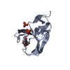
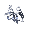
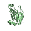
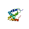
- PDB-6fbk: Crystal structure of the human WNK2 CCT-like 1 domain in complex ... -
+ Open data

- Basic information
| Entry | Database: PDB / ID: 6fbk | ||||||
|---|---|---|---|---|---|---|---|
| Title | Crystal structure of the human WNK2 CCT-like 1 domain in complex with a WNK1 RFXV peptide | ||||||
 Components Components |
| ||||||
 Keywords Keywords | PEPTIDE BINDING PROTEIN / WNK2 / CCT Domain / AI domain / RFXV motif binding | ||||||
| Function / homology |  Function and homology information Function and homology informationpositive regulation of sodium ion transmembrane transporter activity / negative regulation of cell-cell adhesion mediated by integrin / monoatomic cation homeostasis / chemokine (C-C motif) ligand 21 signaling pathway / lymphocyte migration into lymph node / positive regulation of termination of RNA polymerase II transcription / negative regulation of pancreatic juice secretion / protein insertion into ER membrane by stop-transfer membrane-anchor sequence / negative regulation of small GTPase mediated signal transduction / negative regulation of sodium ion transport ...positive regulation of sodium ion transmembrane transporter activity / negative regulation of cell-cell adhesion mediated by integrin / monoatomic cation homeostasis / chemokine (C-C motif) ligand 21 signaling pathway / lymphocyte migration into lymph node / positive regulation of termination of RNA polymerase II transcription / negative regulation of pancreatic juice secretion / protein insertion into ER membrane by stop-transfer membrane-anchor sequence / negative regulation of small GTPase mediated signal transduction / negative regulation of sodium ion transport / monoatomic ion homeostasis / positive regulation of mitotic cytokinesis / intracellular membraneless organelle / negative regulation of heterotypic cell-cell adhesion / regulation of mRNA export from nucleus / intracellular chloride ion homeostasis / positive regulation of T cell chemotaxis / regulation of sodium ion transmembrane transport / cellular response to chemokine / negative regulation of leukocyte cell-cell adhesion / potassium ion homeostasis / cellular hyperosmotic response / membraneless organelle assembly / cell volume homeostasis / regulation of monoatomic cation transmembrane transport / positive regulation of systemic arterial blood pressure / protein kinase activator activity / negative regulation of protein localization to plasma membrane / neuron development / phosphatase binding / regulation of sodium ion transport / monoatomic ion transport / negative regulation of protein ubiquitination / negative regulation of autophagy / sodium ion transmembrane transport / molecular condensate scaffold activity / negative regulation of ERK1 and ERK2 cascade / Stimuli-sensing channels / positive regulation of angiogenesis / mitotic spindle / protein autophosphorylation / positive regulation of canonical Wnt signaling pathway / T cell receptor signaling pathway / heart development / protein phosphorylation / protein kinase activity / non-specific serine/threonine protein kinase / intracellular signal transduction / negative regulation of cell population proliferation / protein serine kinase activity / protein serine/threonine kinase activity / DNA damage response / protein kinase binding / signal transduction / protein-containing complex / ATP binding / membrane / nucleus / plasma membrane / cytoplasm / cytosol Similarity search - Function | ||||||
| Biological species |  Homo sapiens (human) Homo sapiens (human) | ||||||
| Method |  X-RAY DIFFRACTION / SYNCHROTRON / MOLECULAR REPLACEMENT / Resolution: 1.743 Å X-RAY DIFFRACTION / SYNCHROTRON / MOLECULAR REPLACEMENT / Resolution: 1.743 Å | ||||||
 Authors Authors | Pinkas, D.M. / Bufton, J.C. / Burgess-Brown, N.A. / Arrowsmith, C.H. / Edwards, A.M. / Bountra, C. / Bullock, A.N. | ||||||
 Citation Citation | Journal: To Be Published Title: Crystal structure of the human WNK2 CCT-like 1 domain in complex with a WNK1 RFXV peptide Authors: Pinkas, D.M. / Bufton, J.C. / Burgess-Brown, N.A. / Arrowsmith, C.H. / Edwards, A.M. / Bountra, C. / Bullock, A. | ||||||
| History |
|
- Structure visualization
| Structure viewer | Molecule: MolmilJmol/JSmol |
|---|
- Downloads & links
-Download
| PDBx/mmCIF format | 6fbk.cif.gz | 33.3 KB | Display | PDBx/mmCIF format |
|---|---|---|---|---|
| PDB format | pdb6fbk.ent.gz | 21 KB | Display | PDB format |
| PDBx/mmJSON format | 6fbk.json.gz | Tree view | PDBx/mmJSON format | |
| Others |  Other downloads Other downloads |
-Validation report
| Arichive directory | https://data.pdbj.org/pub/pdb/validation_reports/fb/6fbkftp://data.pdbj.org/pub/pdb/validation_reports/fb/6fbk | HTTPS FTP |
|---|
-Related structure data
| Related structure data |  6elmS S: Starting model for refinement |
|---|---|
| Similar structure data |


-Links
 PDBj
PDBj- Assembly
| Deposited unit | 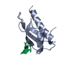
| ||||||||
|---|---|---|---|---|---|---|---|---|---|
| 1 |
| ||||||||
| Unit cell |
|
-Components
| #1: Protein | Mass: 11340.844 Da / Num. of mol.: 1 Source method: isolated from a genetically manipulated source Source: (gene. exp.) Homo sapiens (human) / Gene: WNK2, KIAA1760, PRKWNK2, SDCCAG43, P/OKcl.13 / Production host:  References: UniProt: Q9Y3S1, non-specific serine/threonine protein kinase |
|---|---|
| #2: Protein/peptide | Mass: 2610.046 Da / Num. of mol.: 1 / Source method: obtained synthetically / Source: (synth.) Homo sapiens (human)References: UniProt: Q9H4A3, non-specific serine/threonine protein kinase |
| #3: Water | ChemComp-HOH /  Mass: 18.015 Da / Num. of mol.: 14 / Source method: isolated from a natural source / Formula: H2O Mass: 18.015 Da / Num. of mol.: 14 / Source method: isolated from a natural source / Formula: H2O |
-Experimental details
-Experiment
| Experiment | Method: X-RAY DIFFRACTION / Number of used crystals: 1 |
|---|
- Sample preparation
| Crystal | Density Matthews: 2.18 Å3/Da / Density % sol: 43.45 % |
|---|---|
| Crystal grow | Temperature: 293 K / Method: vapor diffusion / pH: 7.5 Details: 20% PEG3350 -- 10% ethylene glycol -- 0.2M sodium fluoride |
-Data collection
| Diffraction | Mean temperature: 100 K |
|---|---|
| Diffraction source | Source: SYNCHROTRON / Site: Diamond  / Beamline: I04 / Wavelength: 0.9795 Å / Beamline: I04 / Wavelength: 0.9795 Å |
| Detector | Type: DECTRIS PILATUS 6M-F / Detector: PIXEL / Date: Apr 30, 2017 |
| Radiation | Protocol: SINGLE WAVELENGTH / Monochromatic (M) / Laue (L): M / Scattering type: x-ray |
| Radiation wavelength | Wavelength: 0.9795 Å / Relative weight: 1 |
| Reflection | Resolution: 1.74→55.3 Å / Num. obs: 11689 / % possible obs: 99.3 % / Redundancy: 6.5 % / Net I/σ(I): 20.7 |
| Reflection shell | Resolution: 1.743→1.773 Å |
- Processing
| Software |
| |||||||||||||||||||||||||||||||||||
|---|---|---|---|---|---|---|---|---|---|---|---|---|---|---|---|---|---|---|---|---|---|---|---|---|---|---|---|---|---|---|---|---|---|---|---|---|
| Refinement | Method to determine structure: MOLECULAR REPLACEMENT Starting model: 6ELM Resolution: 1.743→33.973 Å / SU ML: 0.31 / Cross valid method: FREE R-VALUE / σ(F): 1.35 / Phase error: 50.6 Details: Refinement R factors are higher than expected for resolution and diffraction images show diffuse spots between the indexed spots that were not able to be integrated. Nevertheless, the structure appears correct.
| |||||||||||||||||||||||||||||||||||
| Solvent computation | Shrinkage radii: 0.9 Å / VDW probe radii: 1.11 Å | |||||||||||||||||||||||||||||||||||
| Refinement step | Cycle: LAST / Resolution: 1.743→33.973 Å
| |||||||||||||||||||||||||||||||||||
| Refine LS restraints |
| |||||||||||||||||||||||||||||||||||
| LS refinement shell |
|
