 Movie
Movie Controller


+ Open data

- Basic information
| Entry | Database: PDB / ID: 6fb4 | |||||||||
|---|---|---|---|---|---|---|---|---|---|---|
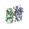
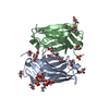
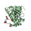
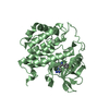
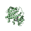
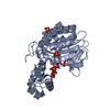
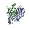
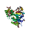
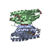
| Title | human KIBRA C2 domain mutant C771A | |||||||||
 Components Components | Protein KIBRA | |||||||||
 Keywords Keywords | LIPID BINDING PROTEIN / C2 domain / Kibra / phosphoinositide-binding / membrane interaction | |||||||||
| Function / homology |  Function and homology information Function and homology informationregulation of hippo signaling / negative regulation of organ growth / hippo signaling / Signaling by Hippo / NOTCH3 Intracellular Domain Regulates Transcription / establishment of cell polarity / negative regulation of hippo signaling / signaling adaptor activity / ruffle membrane / kinase binding ...regulation of hippo signaling / negative regulation of organ growth / hippo signaling / Signaling by Hippo / NOTCH3 Intracellular Domain Regulates Transcription / establishment of cell polarity / negative regulation of hippo signaling / signaling adaptor activity / ruffle membrane / kinase binding / cell migration / molecular adaptor activity / positive regulation of MAPK cascade / transcription coactivator activity / negative regulation of cell population proliferation / regulation of DNA-templated transcription / perinuclear region of cytoplasm / negative regulation of transcription by RNA polymerase II / nucleus / cytoplasm / cytosol Similarity search - Function | |||||||||
| Biological species |  Homo sapiens (human) Homo sapiens (human) | |||||||||
| Method |  X-RAY DIFFRACTION / MOLECULAR REPLACEMENT / Resolution: 2.4156309909 Å X-RAY DIFFRACTION / MOLECULAR REPLACEMENT / Resolution: 2.4156309909 Å | |||||||||
 Authors Authors | Crennell, S.J. / Posner, M.G. / Bagby, S. | |||||||||
| Funding support |  United Kingdom, 1items United Kingdom, 1items
| |||||||||
 Citation Citation | Journal: J. Biol. Chem. / Year: 2018 Title: Distinctive phosphoinositide- and Ca2+-binding properties of normal and cognitive performance-linked variant forms of KIBRA C2 domain. Authors: Posner, M.G. / Upadhyay, A. / Ishima, R. / Kalli, A.C. / Harris, G. / Kremerskothen, J. / Sansom, M.S.P. / Crennell, S.J. / Bagby, S. | |||||||||
| History |
|
- Structure visualization
| Structure viewer | Molecule: MolmilJmol/JSmol |
|---|
- Downloads & links
-Download
| PDBx/mmCIF format | 6fb4.cif.gz | 137.3 KB | Display | PDBx/mmCIF format |
|---|---|---|---|---|
| PDB format | pdb6fb4.ent.gz | 89.2 KB | Display | PDB format |
| PDBx/mmJSON format | 6fb4.json.gz | Tree view | PDBx/mmJSON format | |
| Others |  Other downloads Other downloads |
-Validation report
| Arichive directory | https://data.pdbj.org/pub/pdb/validation_reports/fb/6fb4ftp://data.pdbj.org/pub/pdb/validation_reports/fb/6fb4 | HTTPS FTP |
|---|
-Related structure data
| Related structure data | 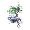 6fd0C  6fjcC 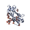 6fjdC  3k9dS S: Starting model for refinement C: citing same article ( |
|---|---|
| Similar structure data |

-Links
 PDBj
PDBj


- Assembly
| Deposited unit | 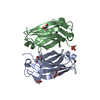
| |||||||||||||||||||||
|---|---|---|---|---|---|---|---|---|---|---|---|---|---|---|---|---|---|---|---|---|---|---|
| 1 |
| |||||||||||||||||||||
| Unit cell |
| |||||||||||||||||||||
| Components on special symmetry positions |
|
-Components
| #1: Protein | Mass: 14547.594 Da / Num. of mol.: 2 / Mutation: C771A Source method: isolated from a genetically manipulated source Details: KIBRA is a multi-functional scaffold protein, the C2 domain binds phosphoinositides. Source: (gene. exp.) Homo sapiens (human) / Gene: WWC1, KIAA0869 / Organ: kidney, brain / Plasmid: pQE30 / Production host:  #2: Chemical | ChemComp-GOL /   Mass: 92.094 Da / Num. of mol.: 4 / Source method: obtained synthetically / Formula: C3H8O3 Mass: 92.094 Da / Num. of mol.: 4 / Source method: obtained synthetically / Formula: C3H8O3#3: Chemical | ChemComp-PO4 / |   Mass: 94.971 Da / Num. of mol.: 1 / Source method: obtained synthetically / Formula: PO4 Mass: 94.971 Da / Num. of mol.: 1 / Source method: obtained synthetically / Formula: PO4#4: Water | ChemComp-HOH / |  Mass: 18.015 Da / Num. of mol.: 124 / Source method: isolated from a natural source / Formula: H2O Mass: 18.015 Da / Num. of mol.: 124 / Source method: isolated from a natural source / Formula: H2OHas protein modification | Y | |
|---|
-Experimental details
-Experiment
| Experiment | Method: X-RAY DIFFRACTION / Number of used crystals: 1 |
|---|
- Sample preparation
| Crystal | Density Matthews: 3.63 Å3/Da / Density % sol: 66.08 % / Description: pavilion |
|---|---|
| Crystal grow | Temperature: 291 K / Method: vapor diffusion, sitting drop / pH: 7.5 Details: 0.1M Na HEPES, 0.8M Na dihydrogen phosphare, 0.8M K dihydrogen phosphate 5% glycerol cryoprotectant |
-Data collection
| Diffraction | Mean temperature: 100 K |
|---|---|
| Diffraction source | Source: ROTATING ANODE / Type: RIGAKU MICROMAX-007 HF / Wavelength: 1.54 Å |
| Detector | Type: RIGAKU SATURN 944+ / Detector: CCD / Date: Sep 3, 2014 |
| Radiation | Protocol: SINGLE WAVELENGTH / Monochromatic (M) / Laue (L): M / Scattering type: x-ray |
| Radiation wavelength | Wavelength: 1.54 Å / Relative weight: 1 |
| Reflection | Resolution: 2.415→72.643 Å / Num. obs: 17355 / % possible obs: 99.9 % / Redundancy: 10.6 % / Biso Wilson estimate: 41.2630665184 Å2 / Rmerge(I) obs: 0.133 / Rrim(I) all: 0.14 / Χ2: 0.96 / Net I/σ(I): 10.7 |
| Reflection shell | Resolution: 2.42→2.5 Å / Redundancy: 6.22 % / Rmerge(I) obs: 0.625 / Mean I/σ(I) obs: 1.7 / Rrim(I) all: 0.676 / Χ2: 1.34 / % possible all: 98.6 |
- Processing
| Software |
| |||||||||||||||||||||||||||||||||||||||||||||||||
|---|---|---|---|---|---|---|---|---|---|---|---|---|---|---|---|---|---|---|---|---|---|---|---|---|---|---|---|---|---|---|---|---|---|---|---|---|---|---|---|---|---|---|---|---|---|---|---|---|---|---|
| Refinement | Method to determine structure: MOLECULAR REPLACEMENT Starting model: 3K9D Resolution: 2.4156309909→72.6422108694 Å / SU ML: 0.325016801318 / Cross valid method: FREE R-VALUE / σ(F): 1.35109357799 / Phase error: 25.3297439476 Stereochemistry target values: GeoStd + Monomer Library + CDL v1.2
| |||||||||||||||||||||||||||||||||||||||||||||||||
| Solvent computation | Shrinkage radii: 0.9 Å / VDW probe radii: 1.11 Å / Solvent model: FLAT BULK SOLVENT MODEL | |||||||||||||||||||||||||||||||||||||||||||||||||
| Displacement parameters | Biso mean: 55.639236193 Å2 | |||||||||||||||||||||||||||||||||||||||||||||||||
| Refinement step | Cycle: LAST / Resolution: 2.4156309909→72.6422108694 Å
| |||||||||||||||||||||||||||||||||||||||||||||||||
| Refine LS restraints |
| |||||||||||||||||||||||||||||||||||||||||||||||||
| LS refinement shell |
|
