 Movie
Movie Controller

[English] 日本語
 Yorodumi
- PDB-6fb1: Crystal Structure of a Tailored I-CreI Homing Endonuclease Protei... -
+ Open data

- Basic information
| Entry | Database: PDB / ID: 6fb1 | ||||||
|---|---|---|---|---|---|---|---|
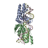
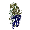
| Title | Crystal Structure of a Tailored I-CreI Homing Endonuclease Protein (3115 variant) in complex with its target DNA (Haemoglobin beta subunit gene) in the presence of Magnesium | ||||||
 Components Components |
| ||||||
 Keywords Keywords | DNA BINDING PROTEIN / Chlamydomonas reinhardtii | ||||||
| Function / homology |  Function and homology information Function and homology informationintron homing / chloroplast / endonuclease activity / Hydrolases; Acting on ester bonds / hydrolase activity / metal ion binding / identical protein binding Similarity search - Function | ||||||
| Biological species |   Chlamydomonas reinhardtii (plant) Chlamydomonas reinhardtii (plant)synthetic construct (others) | ||||||
| Method |  X-RAY DIFFRACTION / SYNCHROTRON / MOLECULAR REPLACEMENT / Resolution: 3.024 Å X-RAY DIFFRACTION / SYNCHROTRON / MOLECULAR REPLACEMENT / Resolution: 3.024 Å | ||||||
 Authors Authors | Molina, R. / Prieto, J. | ||||||
 Citation Citation | Journal: Sci Rep / Year: 2018 Title: Understanding the indirect DNA read-out specificity of I-CreI Meganuclease. Authors: Prieto, J. / Redondo, P. / Lopez-Mendez, B. / D'Abramo, M. / Merino, N. / Blanco, F.J. / Duchateau, P. / Montoya, G. / Molina, R. | ||||||
| History |
|
- Structure visualization
| Structure viewer | Molecule: MolmilJmol/JSmol |
|---|
- Downloads & links
-Download
| PDBx/mmCIF format | 6fb1.cif.gz | 194.4 KB | Display | PDBx/mmCIF format |
|---|---|---|---|---|
| PDB format | pdb6fb1.ent.gz | 151.7 KB | Display | PDB format |
| PDBx/mmJSON format | 6fb1.json.gz | Tree view | PDBx/mmJSON format | |
| Others |  Other downloads Other downloads |
-Validation report
| Arichive directory | https://data.pdbj.org/pub/pdb/validation_reports/fb/6fb1ftp://data.pdbj.org/pub/pdb/validation_reports/fb/6fb1 | HTTPS FTP |
|---|
-Related structure data
| Related structure data |  6fb0C  6fb2C  6fb5C  6fb6C  6fb7C  6fb8C 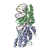 6fb9C  1g9zS S: Starting model for refinement C: citing same article ( |
|---|---|
| Similar structure data |


-Links
 PDBj
PDBj


- Assembly
| Deposited unit | 
| ||||||||
|---|---|---|---|---|---|---|---|---|---|
| 1 |
| ||||||||
| Unit cell |
|
-Components
-DNA endonuclease I- ... , 2 types, 2 molecules AB
| #1: Protein | Mass: 17641.312 Da / Num. of mol.: 1 Source method: isolated from a genetically manipulated source Source: (gene. exp.) Chlamydomonas reinhardtii (plant) / Production host:  References: UniProt: P05725*PLUS, Hydrolases; Acting on ester bonds |
|---|---|
| #2: Protein | Mass: 17583.205 Da / Num. of mol.: 1 Source method: isolated from a genetically manipulated source Source: (gene. exp.) Chlamydomonas reinhardtii (plant) / Production host: References: UniProt: P05725*PLUS, Hydrolases; Acting on ester bonds |
-DNA chain , 4 types, 4 molecules DFEG
| #3: DNA chain | Mass: 4175.730 Da / Num. of mol.: 1 / Source method: obtained synthetically / Source: (synth.) synthetic construct (others) |
|---|---|
| #4: DNA chain | Mass: 4262.764 Da / Num. of mol.: 1 / Source method: obtained synthetically / Source: (synth.) synthetic construct (others) |
| #5: DNA chain | Mass: 3118.067 Da / Num. of mol.: 1 / Source method: obtained synthetically / Source: (synth.) synthetic construct (others) |
| #6: DNA chain | Mass: 3093.054 Da / Num. of mol.: 1 / Source method: obtained synthetically / Source: (synth.) synthetic construct (others) |
-Non-polymers , 2 types, 18 molecules 

| #7: Chemical |  Mass: 24.305 Da / Num. of mol.: 3 / Source method: obtained synthetically / Formula: Mg Mass: 24.305 Da / Num. of mol.: 3 / Source method: obtained synthetically / Formula: Mg#8: Water | ChemComp-HOH / | Mass: 18.015 Da / Num. of mol.: 15 / Source method: isolated from a natural source / Formula: H2O |
|---|
-Experimental details
-Experiment
| Experiment | Method: X-RAY DIFFRACTION / Number of used crystals: 1 |
|---|
- Sample preparation
| Crystal | Density Matthews: 2.75 Å3/Da / Density % sol: 55.28 % |
|---|---|
| Crystal grow | Temperature: 291 K / Method: vapor diffusion, hanging drop Details: 0.1 M calcium acetate 0.1 M sodium acetate pH 4.6 35% (v/v) 1,2-propanediol |
-Data collection
| Diffraction | Mean temperature: 100 K |
|---|---|
| Diffraction source | Source: SYNCHROTRON / Site: SLS  / Beamline: X06SA / Wavelength: 1 Å / Beamline: X06SA / Wavelength: 1 Å |
| Detector | Type: DECTRIS PILATUS 6M / Detector: PIXEL / Date: Nov 16, 2012 |
| Radiation | Protocol: SINGLE WAVELENGTH / Monochromatic (M) / Laue (L): M / Scattering type: x-ray |
| Radiation wavelength | Wavelength: 1 Å / Relative weight: 1 |
| Reflection | Resolution: 3→66.6 Å / Num. obs: 18027 / % possible obs: 99.7 % / Redundancy: 2.4 % / Net I/σ(I): 5.4 |
| Reflection shell | Resolution: 3→3.15 Å |
- Processing
| Software |
| |||||||||||||||||||||||||||||||||||||||||||||||||
|---|---|---|---|---|---|---|---|---|---|---|---|---|---|---|---|---|---|---|---|---|---|---|---|---|---|---|---|---|---|---|---|---|---|---|---|---|---|---|---|---|---|---|---|---|---|---|---|---|---|---|
| Refinement | Method to determine structure: MOLECULAR REPLACEMENT Starting model: 1G9Z Resolution: 3.024→33.985 Å / SU ML: 0.45 / Cross valid method: FREE R-VALUE / σ(F): 1.31 / Phase error: 35.32
| |||||||||||||||||||||||||||||||||||||||||||||||||
| Solvent computation | Shrinkage radii: 0.9 Å / VDW probe radii: 1.11 Å | |||||||||||||||||||||||||||||||||||||||||||||||||
| Refinement step | Cycle: LAST / Resolution: 3.024→33.985 Å
| |||||||||||||||||||||||||||||||||||||||||||||||||
| Refine LS restraints |
| |||||||||||||||||||||||||||||||||||||||||||||||||
| LS refinement shell |
| |||||||||||||||||||||||||||||||||||||||||||||||||
| Refinement TLS params. | Method: refined / Origin x: 6.2003 Å / Origin y: -14.4856 Å / Origin z: 113.9935 Å
| |||||||||||||||||||||||||||||||||||||||||||||||||
| Refinement TLS group | Selection details: all |
