[English] 日本語
Yorodumi
- PDB-6f7t: Crystal Structure of an Fab fragment in complex with a peptide fr... -

+
Open data


ID or keywords:

Loading...

-
Basic information

Entry
Database: PDB / ID: 6f7t
Title
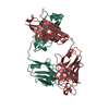
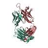
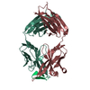
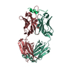
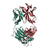
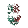
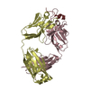
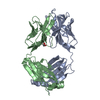
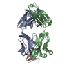
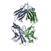
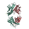
Crystal Structure of an Fab fragment in complex with a peptide from Bacillus subtilis RNase Y
Components
  • FAB RY79-90, HEAVY CHAIN
  • FAB RY79-90, LIGHT CHAIN
  • Ribonuclease Y
KeywordsIMMUNE SYSTEM / Fab-peptide complex / lambda x light chain / RNase Y
Function / homology
Function and homology information


mRNA catabolic process / RNA endonuclease activity / mRNA processing / Hydrolases; Acting on ester bonds / membrane => GO:0016020 / RNA binding / identical protein binding / plasma membrane
Similarity search - Function
Ribonuclease Y / Ribonuclease Y, N-terminal / RNase Y N-terminal region / HDIG domain / KH domain / HD domain profile. / K Homology domain, type 1 / HD domain / HD domain / Type-1 KH domain profile. ...Ribonuclease Y / Ribonuclease Y, N-terminal / RNase Y N-terminal region / HDIG domain / KH domain / HD domain profile. / K Homology domain, type 1 / HD domain / HD domain / Type-1 KH domain profile. / K Homology domain, type 1 superfamily / Metal dependent phosphohydrolases with conserved 'HD' motif. / HD/PDEase domain / K Homology domain / K homology RNA-binding domain / Immunoglobulins / Immunoglobulin-like / Sandwich / Mainly Beta
Similarity search - Domain/homology
Biological speciesMus musculus (house mouse)
Bacillus subtilis (bacteria)
MethodX-RAY DIFFRACTION / SYNCHROTRON / MOLECULAR REPLACEMENT / Resolution: 2.6 Å
AuthorsGolinelli-Pimpaneau, B. / Hardouin, P.
Funding support France, 2items
OrganizationGrant numberCountry
LABEX DynamoANR-11-LABX-0011 France
French National Research AgencyANR-11-BSV8-026 France
CitationJournal: Biophys. J. / Year: 2018
Title: Dissociation of the Dimer of the Intrinsically Disordered Domain of RNase Y upon Antibody Binding.
Authors: Hardouin, P. / Velours, C. / Bou-Nader, C. / Assrir, N. / Laalami, S. / Putzer, H. / Durand, D. / Golinelli-Pimpaneau, B.
History
DepositionDec 11, 2017Deposition site: PDBE / Processing site: PDBE
Revision 1.0Oct 31, 2018Provider: repository / Type: Initial release
Revision 1.1Nov 28, 2018Group: Data collection / Database references / Category: citation
Item: _citation.country / _citation.journal_abbrev ..._citation.country / _citation.journal_abbrev / _citation.journal_id_ASTM / _citation.journal_id_CSD / _citation.journal_id_ISSN / _citation.pdbx_database_id_DOI / _citation.pdbx_database_id_PubMed / _citation.title / _citation.year
Revision 1.2Dec 19, 2018Group: Data collection / Database references / Category: citation
Item: _citation.journal_volume / _citation.page_first / _citation.page_last
Revision 1.3Jan 17, 2024Group: Data collection / Database references / Refinement description
Category: chem_comp_atom / chem_comp_bond ...chem_comp_atom / chem_comp_bond / database_2 / pdbx_initial_refinement_model
Item: _database_2.pdbx_DOI / _database_2.pdbx_database_accession

-
Structure visualization

Structure viewerMolecule:
MolmilJmol/JSmol

Downloads & links

-
Assembly

Deposited unit
A: FAB RY79-90, LIGHT CHAIN
B: FAB RY79-90, HEAVY CHAIN
C: Ribonuclease Y
D: Ribonuclease Y
H: FAB RY79-90, HEAVY CHAIN
L: FAB RY79-90, LIGHT CHAIN
hetero molecules


Theoretical massNumber of molelcules
Total (without water)96,0977
Polymers96,0736
Non-polymers241
Water4,648258
1
A: FAB RY79-90, LIGHT CHAIN
B: FAB RY79-90, HEAVY CHAIN
D: Ribonuclease Y


Theoretical massNumber of molelcules
Total (without water)48,0373
Polymers48,0373
Non-polymers00
Water543
TypeNameSymmetry operationNumber
identity operation1_555x,y,z1
Buried area4940 Å2
ΔGint-28 kcal/mol
Surface area20630 Å2
MethodPISA
2
C: Ribonuclease Y
H: FAB RY79-90, HEAVY CHAIN
L: FAB RY79-90, LIGHT CHAIN
hetero molecules


Theoretical massNumber of molelcules
Total (without water)48,0614
Polymers48,0373
Non-polymers241
Water543
TypeNameSymmetry operationNumber
identity operation1_555x,y,z1
Buried area5020 Å2
ΔGint-27 kcal/mol
Surface area20680 Å2
MethodPISA
Unit cell
Length a, b, c (Å)98.680, 130.450, 72.050
Angle α, β, γ (deg.)90.00, 90.00, 90.00
Int Tables number18
Space group name H-MP21212
Components on special symmetry positions
IDModelComponents
11L-301-

MG

21L-430-

HOH

-
Components

#1: Antibody FAB RY79-90, LIGHT CHAIN


Mass: 23633.234 Da / Num. of mol.: 2 / Source method: isolated from a natural source / Source: (natural) Mus musculus (house mouse)
#2: Antibody FAB RY79-90, HEAVY CHAIN


Mass: 22799.588 Da / Num. of mol.: 2 / Source method: isolated from a natural source / Source: (natural) Mus musculus (house mouse)
#3: Protein/peptide Ribonuclease Y / RNase Y


Mass: 1603.739 Da / Num. of mol.: 2 / Fragment: UNP residues 79-90 / Source method: obtained synthetically / Source: (synth.) Bacillus subtilis (bacteria)
References: UniProt: O31774, Hydrolases; Acting on ester bonds
#4: Chemical ChemComp-MG / MAGNESIUM ION


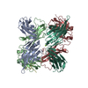
Mass: 24.305 Da / Num. of mol.: 1 / Source method: obtained synthetically / Formula: Mg
#5: Water ChemComp-HOH / water / Water


Mass: 18.015 Da / Num. of mol.: 258 / Source method: isolated from a natural source / Formula: H2O

-
Experimental details

-
Experiment

ExperimentMethod: X-RAY DIFFRACTION / Number of used crystals: 1

-
Sample preparation

CrystalDensity Matthews: 2.42 Å3/Da / Density % sol: 49.1 %
Crystal growTemperature: 298 K / Method: vapor diffusion, hanging drop / pH: 8.5 / Details: 0.2M MgCl2; 0.1M TRIS; 16% (w/v) PEG 4000

-
Data collection

DiffractionMean temperature: 80 K
Diffraction sourceSource: SYNCHROTRON / Site: SOLEIL / Beamline: PROXIMA 1 / Wavelength: 0.97934 Å
DetectorType: DECTRIS PILATUS 6M / Detector: PIXEL / Date: Mar 19, 2016
RadiationProtocol: SINGLE WAVELENGTH / Monochromatic (M) / Laue (L): M / Scattering type: x-ray
Radiation wavelengthWavelength: 0.97934 Å / Relative weight: 1
ReflectionResolution: 2.6→50 Å / Num. obs: 29393 / % possible obs: 99.7 % / Redundancy: 8.7 % / Biso Wilson estimate: 84.31 Å2 / CC1/2: 0.998 / Net I/σ(I): 14.6
Reflection shellResolution: 2.6→2.75 Å / Redundancy: 8.63 % / Mean I/σ(I) obs: 1.41 / CC1/2: 0.586 / % possible all: 98.2

-
Processing

Software
NameVersionClassification
BUSTER2.10.3refinement
XDS0.7.14data reduction
XSCALEdata scaling
PHASERphasing
RefinementMethod to determine structure: MOLECULAR REPLACEMENT
Starting model: 2QHR
Resolution: 2.6→46.15 Å / Cor.coef. Fo:Fc: 0.946 / Cor.coef. Fo:Fc free: 0.927 / SU R Cruickshank DPI: 1.249 / Cross valid method: THROUGHOUT / σ(F): 0 / SU R Blow DPI: 2.905 / SU Rfree Blow DPI: 0.321 / SU Rfree Cruickshank DPI: 0.321
RfactorNum. reflection% reflectionSelection details
Rfree0.253 1464 5 %RANDOM
Rwork0.202 ---
obs0.204 29299 100 %-
Displacement parametersBiso mean: 84.68 Å2
Baniso -1Baniso -2Baniso -3
1--2.8445 Å20 Å20 Å2
2---4.3245 Å20 Å2
3---7.169 Å2
Refine analyzeLuzzati coordinate error obs: 0.36 Å
Refinement stepCycle: 1 / Resolution: 2.6→46.15 Å
ProteinNucleic acidLigandSolventTotal
Num. atoms6741 0 1 258 7000
Refine LS restraints
Refine-IDTypeDev idealNumberRestraint functionWeight
X-RAY DIFFRACTIONt_bond_d0.016929HARMONIC2
X-RAY DIFFRACTIONt_angle_deg1.269441HARMONIC2
X-RAY DIFFRACTIONt_dihedral_angle_d2303SINUSOIDAL2
X-RAY DIFFRACTIONt_incorr_chiral_ct
X-RAY DIFFRACTIONt_pseud_angle
X-RAY DIFFRACTIONt_trig_c_planes
X-RAY DIFFRACTIONt_gen_planes1153HARMONIC5
X-RAY DIFFRACTIONt_it6929HARMONIC20
X-RAY DIFFRACTIONt_nbd0SEMIHARMONIC5
X-RAY DIFFRACTIONt_omega_torsion3.2
X-RAY DIFFRACTIONt_other_torsion21.19
X-RAY DIFFRACTIONt_improper_torsion
X-RAY DIFFRACTIONt_chiral_improper_torsion942SEMIHARMONIC5
X-RAY DIFFRACTIONt_sum_occupancies
X-RAY DIFFRACTIONt_utility_distance
X-RAY DIFFRACTIONt_utility_angle
X-RAY DIFFRACTIONt_utility_torsion
X-RAY DIFFRACTIONt_ideal_dist_contact7606SEMIHARMONIC4
LS refinement shellResolution: 2.6→2.69 Å / Total num. of bins used: 15
RfactorNum. reflection% reflection
Rfree0.2881 141 4.99 %
Rwork0.2261 2684 -
all0.2292 2825 -
obs--100 %
Refinement TLS params.

Method: refined / Refine-ID: X-RAY DIFFRACTION

IDL112)L122)L132)L222)L232)L332)S11 (Å °)S12 (Å °)S13 (Å °)S21 (Å °)S22 (Å °)S23 (Å °)S31 (Å °)S32 (Å °)S33 (Å °)T112)T122)T132)T222)T232)T332)Origin x (Å)Origin y (Å)Origin z (Å)
11.35910.0260.21260.6595-0.12290.77530.08110.034-0.08-0.0017-0.0137-0.03490.0189-0.2937-0.0674-0.22690.0420.0067-0.00010.0083-0.2262-17.3109-2.8843-43.5537
20.33390.14880.42480.6565-0.41660.17490.021-0.1295-0.0688-0.07530.05520.21750.0064-0.3305-0.0763-0.18760.0459-0.01970.08440.0197-0.0647-32.5924-1.4173-45.327
Refinement TLS group
IDRefine-IDRefine TLS-IDSelection details
1X-RAY DIFFRACTION1{ A|* L|* C|*}
2X-RAY DIFFRACTION2{ B|* H|* D|*}

+
About Yorodumi

-
News

-
Feb 9, 2022. New format data for meta-information of EMDB entries

New format data for meta-information of EMDB entries

  • Version 3 of the EMDB header file is now the official format.
  • The previous official version 1.9 will be removed from the archive.

Related info.:EMDB header

External links:wwPDB to switch to version 3 of the EMDB data model

-
Aug 12, 2020. Covid-19 info

Covid-19 info

URL: https://pdbj.org/emnavi/covid19.php

New page: Covid-19 featured information page in EM Navigator.

Related info.:Covid-19 info / Mar 5, 2020. Novel coronavirus structure data

+
Mar 5, 2020. Novel coronavirus structure data

Novel coronavirus structure data

Related info.:Yorodumi Speices / Aug 12, 2020. Covid-19 info

External links:COVID-19 featured content - PDBj / Molecule of the Month (242):Coronavirus Proteases

+
Jan 31, 2019. EMDB accession codes are about to change! (news from PDBe EMDB page)

EMDB accession codes are about to change! (news from PDBe EMDB page)

  • The allocation of 4 digits for EMDB accession codes will soon come to an end. Whilst these codes will remain in use, new EMDB accession codes will include an additional digit and will expand incrementally as the available range of codes is exhausted. The current 4-digit format prefixed with “EMD-” (i.e. EMD-XXXX) will advance to a 5-digit format (i.e. EMD-XXXXX), and so on. It is currently estimated that the 4-digit codes will be depleted around Spring 2019, at which point the 5-digit format will come into force.
  • The EM Navigator/Yorodumi systems omit the EMD- prefix.

Related info.:Q: What is EMD? / ID/Accession-code notation in Yorodumi/EM Navigator

External links:EMDB Accession Codes are Changing Soon! / Contact to PDBj

+
Jul 12, 2017. Major update of PDB

Major update of PDB

  • wwPDB released updated PDB data conforming to the new PDBx/mmCIF dictionary.
  • This is a major update changing the version number from 4 to 5, and with Remediation, in which all the entries are updated.
  • In this update, many items about electron microscopy experimental information are reorganized (e.g. em_software).
  • Now, EM Navigator and Yorodumi are based on the updated data.

External links:wwPDB Remediation / Enriched Model Files Conforming to OneDep Data Standards Now Available in the PDB FTP Archive

-
Yorodumi

Thousand views of thousand structures

  • Yorodumi is a browser for structure data from EMDB, PDB, SASBDB, etc.
  • This page is also the successor to EM Navigator detail page, and also detail information page/front-end page for Omokage search.
  • The word "yorodu" (or yorozu) is an old Japanese word meaning "ten thousand". "mi" (miru) is to see.

Related info.:EMDB / PDB / SASBDB / Comparison of 3 databanks / Yorodumi Search / Aug 31, 2016. New EM Navigator & Yorodumi / Yorodumi Papers / Jmol/JSmol / Function and homology information / Changes in new EM Navigator and Yorodumi

Read more