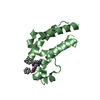
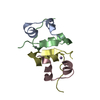
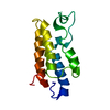
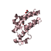
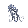
polysaccharide binding / polysaccharide catabolic process / hydrolase activity, hydrolyzing O-glycosyl compounds / metal ion binding Similarity search - Function
#1: Journal: Biomol NMR Assign / Year: 2017 Title: Chemical shift assignments for the apo-form of the catalytic domain, the linker region, and the carbohydrate-binding domain of the cellulose-active lytic polysaccharide monooxygenase ScLPMO10C. Authors: Courtade, G. / Forsberg, Z. / Vaaje-Kolstad, G. / Eijsink, V.G.H. / Aachmann, F.L.
History
Deposition
Dec 8, 2017
Deposition site: PDBE / Processing site: PDBE
Revision 1.0
Jul 11, 2018
Provider: repository / Type: Initial release
Revision 1.1
May 8, 2019
Group: Data collection / Category: pdbx_nmr_software / Item: _pdbx_nmr_software.name
In the structure databanks used in Yorodumi, some data are registered as the other names, "COVID-19 virus" and "2019-nCoV". Here are the details of the virus and the list of structure data.
Jan 31, 2019. EMDB accession codes are about to change! (news from PDBe EMDB page)
EMDB accession codes are about to change! (news from PDBe EMDB page)
The allocation of 4 digits for EMDB accession codes will soon come to an end. Whilst these codes will remain in use, new EMDB accession codes will include an additional digit and will expand incrementally as the available range of codes is exhausted. The current 4-digit format prefixed with “EMD-” (i.e. EMD-XXXX) will advance to a 5-digit format (i.e. EMD-XXXXX), and so on. It is currently estimated that the 4-digit codes will be depleted around Spring 2019, at which point the 5-digit format will come into force.
The EM Navigator/Yorodumi systems omit the EMD- prefix.
Related info.:Q: What is EMD? / ID/Accession-code notation in Yorodumi/EM Navigator
Yorodumi is a browser for structure data from EMDB, PDB, SASBDB, etc.
This page is also the successor to EM Navigator detail page, and also detail information page/front-end page for Omokage search.
The word "yorodu" (or yorozu) is an old Japanese word meaning "ten thousand". "mi" (miru) is to see.
Related info.:EMDB / PDB / SASBDB / Comparison of 3 databanks / Yorodumi Search / Aug 31, 2016. New EM Navigator & Yorodumi / Yorodumi Papers / Jmol/JSmol / Function and homology information / Changes in new EM Navigator and Yorodumi
 Movie
Movie Controller

 Yorodumi
 Open data
 Basic information
 Components
 Keywords
 Function and homology information
 Authors
 Norway, 2items
  Citation
 Structure visualization
 Downloads & links
 Other downloads
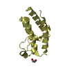


 PDBj
PDBj
 Assembly
 Streptomyces coelicolor A3(2) (bacteria)
 HSQC
HSQC Sample preparation
 Processing