 Movie
Movie Controller

[English] 日本語
 Yorodumi
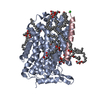
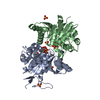
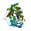
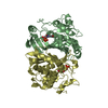
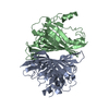
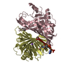
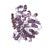
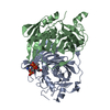
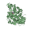
- PDB-6f34: Crystal structure of a bacterial cationic amino acid transporter ... -
+ Open data

- Basic information
| Entry | Database: PDB / ID: 6f34 | ||||||
|---|---|---|---|---|---|---|---|
| Title | Crystal structure of a bacterial cationic amino acid transporter (CAT) homologue bound to Arginine. | ||||||
 Components Components |
| ||||||
 Keywords Keywords | MEMBRANE PROTEIN / Amino Acid Transporter / SLC7 | ||||||
| Function / homology |  Function and homology information Function and homology informationmagnesium ion homeostasis / cellular response to magnesium starvation / amino acid transmembrane transporter activity / membrane / plasma membrane Similarity search - Function | ||||||
| Biological species |  Geobacillus kaustophilus (bacteria) Geobacillus kaustophilus (bacteria) | ||||||
| Method |  X-RAY DIFFRACTION / SYNCHROTRON / MOLECULAR REPLACEMENT / Resolution: 3.13 Å X-RAY DIFFRACTION / SYNCHROTRON / MOLECULAR REPLACEMENT / Resolution: 3.13 Å | ||||||
 Authors Authors | Jungnickel, K.E.J. / Newstead, S. | ||||||
| Funding support |  United Kingdom, 1items United Kingdom, 1items
| ||||||
 Citation Citation | Journal: Nat Commun / Year: 2018 Title: Structural basis for amino acid transport by the CAT family of SLC7 transporters. Authors: Jungnickel, K.E.J. / Parker, J.L. / Newstead, S. | ||||||
| History |
|
- Structure visualization
| Structure viewer | Molecule: MolmilJmol/JSmol |
|---|
- Downloads & links
-Download
| PDBx/mmCIF format | 6f34.cif.gz | 207.3 KB | Display | PDBx/mmCIF format |
|---|---|---|---|---|
| PDB format | pdb6f34.ent.gz | 167.2 KB | Display | PDB format |
| PDBx/mmJSON format | 6f34.json.gz | Tree view | PDBx/mmJSON format | |
| Others |  Other downloads Other downloads |
-Validation report
| Arichive directory | https://data.pdbj.org/pub/pdb/validation_reports/f3/6f34ftp://data.pdbj.org/pub/pdb/validation_reports/f3/6f34 | HTTPS FTP |
|---|
-Related structure data
| Related structure data |  5oqtSC S: Starting model for refinement C: citing same article ( |
|---|---|
| Similar structure data |

-Links
 PDBj
PDBj


- Assembly
| Deposited unit | 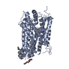
| ||||||||
|---|---|---|---|---|---|---|---|---|---|
| 1 |
| ||||||||
| Unit cell |
|
-Components
-Protein / Protein/peptide , 2 types, 2 molecules AC
| #1: Protein | Mass: 49658.531 Da / Num. of mol.: 1 Source method: isolated from a genetically manipulated source Source: (gene. exp.) Geobacillus kaustophilus (bacteria) / Strain: HTA426 / Gene: GK0930, ApcT / Production host: |
|---|---|
| #2: Protein/peptide | Mass: 2965.577 Da / Num. of mol.: 1 Source method: isolated from a genetically manipulated source Source: (gene. exp.) Strain: K12 / Gene: yneM, b4599, JW1527.1 / Production host: |
-Non-polymers , 4 types, 78 molecules 


| #3: Chemical | ChemComp-ARG /  Type: L-peptide linking / Mass: 175.209 Da / Num. of mol.: 1 / Source method: obtained synthetically / Formula: C6H15N4O2 Type: L-peptide linking / Mass: 175.209 Da / Num. of mol.: 1 / Source method: obtained synthetically / Formula: C6H15N4O2 | ||
|---|---|---|---|
| #4: Chemical | ChemComp-CLR /  Mass: 386.654 Da / Num. of mol.: 1 / Source method: obtained synthetically / Formula: C27H46O Mass: 386.654 Da / Num. of mol.: 1 / Source method: obtained synthetically / Formula: C27H46O | ||
| #5: Chemical |  Mass: 356.540 Da / Num. of mol.: 2 / Source method: obtained synthetically / Formula: C21H40O4 Mass: 356.540 Da / Num. of mol.: 2 / Source method: obtained synthetically / Formula: C21H40O4#6: Water | ChemComp-HOH / | Mass: 18.015 Da / Num. of mol.: 74 / Source method: isolated from a natural source / Formula: H2O |
-Experimental details
-Experiment
| Experiment | Method: X-RAY DIFFRACTION / Number of used crystals: 1 |
|---|
- Sample preparation
| Crystal | Density Matthews: 3.62 Å3/Da / Density % sol: 65.99 % |
|---|---|
| Crystal grow | Temperature: 292 K / Method: lipidic cubic phase / pH: 4 Details: 28-34 % PEG 400, 0.1 M sodium acetate pH 4.0 and 0.1 M potassium fluoride, containing 10 mM of the amino acid ligand. |
-Data collection
| Diffraction | Mean temperature: 77 K | ||||||||||||||||||||||||
|---|---|---|---|---|---|---|---|---|---|---|---|---|---|---|---|---|---|---|---|---|---|---|---|---|---|
| Diffraction source | Source: SYNCHROTRON / Site: SOLEIL  / Beamline: PROXIMA 2 / Wavelength: 0.98 Å / Beamline: PROXIMA 2 / Wavelength: 0.98 Å | ||||||||||||||||||||||||
| Detector | Type: DECTRIS EIGER X 9M / Detector: PIXEL / Date: Sep 30, 2017 | ||||||||||||||||||||||||
| Radiation | Protocol: SINGLE WAVELENGTH / Monochromatic (M) / Laue (L): M / Scattering type: x-ray | ||||||||||||||||||||||||
| Radiation wavelength | Wavelength: 0.98 Å / Relative weight: 1 | ||||||||||||||||||||||||
| Reflection | Resolution: 3.13→64.63 Å / Num. obs: 13951 / % possible obs: 99.9 % / Redundancy: 6.4 % / Biso Wilson estimate: 94.64 Å2 / CC1/2: 0.998 / Rmerge(I) obs: 0.173 / Rpim(I) all: 0.074 / Rrim(I) all: 0.188 / Net I/σ(I): 5.5 / Num. measured all: 89902 / Scaling rejects: 84 | ||||||||||||||||||||||||
| Reflection shell | Diffraction-ID: 1
|
- Processing
| Software |
| |||||||||||||||||||||||||||||||||||||||||||||||||
|---|---|---|---|---|---|---|---|---|---|---|---|---|---|---|---|---|---|---|---|---|---|---|---|---|---|---|---|---|---|---|---|---|---|---|---|---|---|---|---|---|---|---|---|---|---|---|---|---|---|---|
| Refinement | Method to determine structure: MOLECULAR REPLACEMENT Starting model: 5OQT Resolution: 3.13→50.926 Å / SU ML: 0.46 / Cross valid method: THROUGHOUT / σ(F): 1.34 / Phase error: 32.62
| |||||||||||||||||||||||||||||||||||||||||||||||||
| Solvent computation | Shrinkage radii: 0.9 Å / VDW probe radii: 1.11 Å | |||||||||||||||||||||||||||||||||||||||||||||||||
| Displacement parameters | Biso max: 215.42 Å2 / Biso mean: 99.082 Å2 / Biso min: 55.19 Å2 | |||||||||||||||||||||||||||||||||||||||||||||||||
| Refinement step | Cycle: final / Resolution: 3.13→50.926 Å
| |||||||||||||||||||||||||||||||||||||||||||||||||
| Refine LS restraints |
| |||||||||||||||||||||||||||||||||||||||||||||||||
| LS refinement shell | Refine-ID: X-RAY DIFFRACTION / Rfactor Rfree error: 0 / Total num. of bins used: 6
| |||||||||||||||||||||||||||||||||||||||||||||||||
| Refinement TLS params. | Method: refined / Origin x: -12.3337 Å / Origin y: -1.9101 Å / Origin z: 25.4411 Å
| |||||||||||||||||||||||||||||||||||||||||||||||||
| Refinement TLS group |
|
