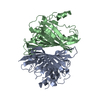
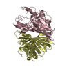
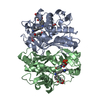
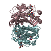
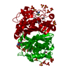
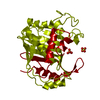
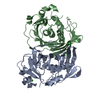
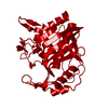
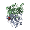
Entry Database : PDB / ID : 4r4uTitle Crystal structure of acyl-CoA thioesterase tesB from Yersinia pestis in complex with coenzyme A Acyl-CoA thioesterase II Keywords / / / / Function / homology Function Domain/homology Component
/ / / / / / / / / / Biological species Yersinia pestis (bacteria)Method / / / Resolution : 2.2 Å Authors Swarbrick, C.M.D. / Forwood, J.K. Journal : Acta Crystallogr.,Sect.D / Year : 2015Title : Structural and functional characterization of TesB from Yersinia pestis reveals a unique octameric arrangement of hotdog domains.Authors : Swarbrick, C.M. / Perugini, M.A. / Cowieson, N. / Forwood, J.K. History Deposition Aug 19, 2014 Deposition site / Processing site Revision 1.0 Apr 15, 2015 Provider / Type Revision 1.1 Apr 29, 2015 Group Revision 1.2 Jul 17, 2019 Group / Refinement description / Category Item / _software.name / _software.versionRevision 1.3 Sep 20, 2023 Group Data collection / Database references ... Data collection / Database references / Derived calculations / Refinement description Category chem_comp_atom / chem_comp_bond ... chem_comp_atom / chem_comp_bond / database_2 / pdbx_initial_refinement_model / pdbx_struct_conn_angle / struct_conn / struct_ref_seq_dif / struct_site Item _database_2.pdbx_DOI / _database_2.pdbx_database_accession ... _database_2.pdbx_DOI / _database_2.pdbx_database_accession / _pdbx_struct_conn_angle.ptnr1_auth_asym_id / _pdbx_struct_conn_angle.ptnr1_auth_seq_id / _pdbx_struct_conn_angle.ptnr1_label_asym_id / _pdbx_struct_conn_angle.ptnr2_auth_asym_id / _pdbx_struct_conn_angle.ptnr2_auth_seq_id / _pdbx_struct_conn_angle.ptnr2_label_asym_id / _pdbx_struct_conn_angle.ptnr3_auth_asym_id / _pdbx_struct_conn_angle.ptnr3_auth_seq_id / _pdbx_struct_conn_angle.ptnr3_label_asym_id / _pdbx_struct_conn_angle.value / _struct_conn.pdbx_dist_value / _struct_conn.ptnr1_auth_asym_id / _struct_conn.ptnr1_auth_comp_id / _struct_conn.ptnr1_auth_seq_id / _struct_conn.ptnr1_label_asym_id / _struct_conn.ptnr1_label_atom_id / _struct_conn.ptnr1_label_comp_id / _struct_conn.ptnr1_label_seq_id / _struct_conn.ptnr2_auth_asym_id / _struct_conn.ptnr2_auth_comp_id / _struct_conn.ptnr2_auth_seq_id / _struct_conn.ptnr2_label_asym_id / _struct_conn.ptnr2_label_atom_id / _struct_conn.ptnr2_label_comp_id / _struct_ref_seq_dif.details / _struct_site.pdbx_auth_asym_id / _struct_site.pdbx_auth_comp_id / _struct_site.pdbx_auth_seq_id
Show all Show less
 Movie
Movie Controller

 Yorodumi
 Open data
 Basic information
 Components
 Keywords
 Function and homology information
 Yersinia pestis (bacteria)
 X-RAY DIFFRACTION /
  Authors
 Citation
 Structure visualization
 Downloads & links
 Other downloads


 PDBj
PDBj


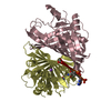
 Assembly

 Mass: 22.990 Da / Num. of mol.: 2 / Source method: obtained synthetically / Formula: Na
 Mass: 767.534 Da / Num. of mol.: 2 / Source method: obtained synthetically / Formula: C21H36N7O16P3S
 Mass: 104.061 Da / Num. of mol.: 1 / Source method: obtained synthetically / Formula: C3H4O4
 Mass: 18.015 Da / Num. of mol.: 414 / Source method: isolated from a natural source / Formula: H2O
 Sample preparation
 / Beamline: MX1 / Wavelength: 0.9537 Å
 Processing