[English] 日本語
Yorodumi- PDB-6ezi: PDZK1 domain 4 in complex with C-terminal peptide of human PepT2. -
+Open data
-Basic information
Entry | Database: PDB / ID: 6ezi | ||||||
---|---|---|---|---|---|---|---|
Title | PDZK1 domain 4 in complex with C-terminal peptide of human PepT2. | ||||||
Components |
| ||||||
Keywords | PROTEIN BINDING / PDZK1 Human peptide transporter Na(+)-H(+) exchange regulatory cofactor NHE-RF3 Protein complex binding | ||||||
Function / homology | Function and homology information positive regulation of cation transmembrane transport / carnitine transport / tripeptide import across plasma membrane / positive regulation of ion transmembrane transporter activity / Proton/oligopeptide cotransporters / peptidoglycan transport / tripeptide transmembrane transporter activity / dipeptide transport / dipeptide import across plasma membrane / peptide:proton symporter activity ...positive regulation of cation transmembrane transport / carnitine transport / tripeptide import across plasma membrane / positive regulation of ion transmembrane transporter activity / Proton/oligopeptide cotransporters / peptidoglycan transport / tripeptide transmembrane transporter activity / dipeptide transport / dipeptide import across plasma membrane / peptide:proton symporter activity / dipeptide transmembrane transporter activity / scavenger receptor binding / antibacterial innate immune response / regulation of nucleotide-binding domain, leucine rich repeat containing receptor signaling pathway / xenobiotic detoxification by transmembrane export across the plasma membrane / positive regulation of protein localization to membrane / xenobiotic transport / peptide transport / microvillus membrane / renal absorption / brush border / transport across blood-brain barrier / positive regulation of protein targeting to membrane / monoatomic ion transport / protein-membrane adaptor activity / regulation of monoatomic anion transport / protein localization to plasma membrane / PDZ domain binding / brush border membrane / phagocytic vesicle membrane / protein transport / apical plasma membrane / signaling receptor binding / protein-containing complex binding / extracellular exosome / plasma membrane Similarity search - Function | ||||||
Biological species | Homo sapiens (human) | ||||||
Method | X-RAY DIFFRACTION / SYNCHROTRON / MOLECULAR REPLACEMENT / Resolution: 1.50332103817 Å | ||||||
Authors | Loew, C. / Flayhan, A. / Pieprzyk, J. | ||||||
Citation | 
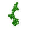
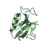
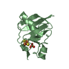
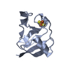
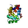
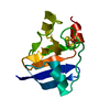
Journal: Structure / Year: 2018 Title: Probing the Architecture of a Multi-PDZ Domain Protein: Structure of PDZK1 in Solution. Authors: Nelly R Hajizadeh / Joanna Pieprzyk / Petr Skopintsev / Ali Flayhan / Dmitri I Svergun / Christian Löw / Abstract: The scaffolding protein PDZK1 has been associated with the regulation of membrane transporters. It contains four conserved PDZ domains, which typically recognize a 3-5-residue long motif at the C ...The scaffolding protein PDZK1 has been associated with the regulation of membrane transporters. It contains four conserved PDZ domains, which typically recognize a 3-5-residue long motif at the C terminus of the binding partner. The atomic structures of the individual domains are available but their spatial arrangement in the full-length context influencing the binding properties remained elusive. Here we report a systematic study of full-length PDZK1 and deletion constructs using small-angle X-ray scattering, complemented with biochemical and functional studies on PDZK1 binding to known membrane protein partners. A hybrid modeling approach utilizing multiple scattering datasets yielded a well-defined, extended, asymmetric L-shaped domain organization of PDZK1 in contrast to a flexible "beads-on-string" model predicted by bioinformatics analysis. The linker regions of PDZK1 appear to play a central role in the arrangement of the four domains underlying the importance of studying scaffolding proteins in their full-length context. | ||||||
History |
|
-Structure visualization
Structure viewer | Molecule: MolmilJmol/JSmol |
---|
-Downloads & links
-Download
PDBx/mmCIF format | 6ezi.cif.gz | 63.5 KB | Display | PDBx/mmCIF format |
---|---|---|---|---|
PDB format | pdb6ezi.ent.gz | 41.7 KB | Display | PDB format |
PDBx/mmJSON format | 6ezi.json.gz | Tree view | PDBx/mmJSON format | |
Others | Other downloads |
-Validation report
Summary document | 6ezi_validation.pdf.gz | 431.5 KB | Display | wwPDB validaton report |
---|---|---|---|---|
Full document | 6ezi_full_validation.pdf.gz | 432.2 KB | Display | |
Data in XML | 6ezi_validation.xml.gz | 7.7 KB | Display | |
Data in CIF | 6ezi_validation.cif.gz | 10.1 KB | Display | |
Arichive directory | https://data.pdbj.org/pub/pdb/validation_reports/ez/6ezi ftp://data.pdbj.org/pub/pdb/validation_reports/ez/6ezi | HTTPS FTP |
-Related structure data
Related structure data | 4r2zS S: Starting model for refinement C: citing same article (ref.) |
---|---|
Similar structure data |
-Links
-Assembly
Deposited unit |
| ||||||||||
---|---|---|---|---|---|---|---|---|---|---|---|
1 |
| ||||||||||
Unit cell |
|
-Components
#1: Protein | Mass: 9653.086 Da / Num. of mol.: 1 Source method: isolated from a genetically manipulated source Source: (gene. exp.) Homo sapiens (human) / Gene: PDZK1, CAP70, NHERF3, PDZD1 / Production host: Escherichia coli BL21(DE3) (bacteria) / References: UniProt: Q5T2W1 |
---|---|
#2: Protein/peptide | Mass: 1205.529 Da / Num. of mol.: 1 / Source method: obtained synthetically / Source: (synth.) Homo sapiens (human) / References: UniProt: Q16348 |
#3: Chemical | ChemComp-GOL / |
#4: Water | ChemComp-HOH / |
-Experimental details
-Experiment
Experiment | Method: X-RAY DIFFRACTION / Number of used crystals: 1 |
---|
-Sample preparation
Crystal | Density Matthews: 2.98 Å3/Da / Density % sol: 58.66 % |
---|---|
Crystal grow | Temperature: 292.15 K / Method: vapor diffusion, sitting drop / pH: 7.5 Details: 0.1 M Trisodium citrate pH 6.45 20% PEG 4000 20 %(v/v) isopropanol |
-Data collection
Diffraction | Mean temperature: 173 K |
---|---|
Diffraction source | Source: SYNCHROTRON / Site: ESRF / Beamline: ID30B / Wavelength: 1.07166 Å |
Detector | Type: DECTRIS PILATUS3 6M / Detector: PIXEL / Date: Feb 13, 2017 |
Radiation | Protocol: SINGLE WAVELENGTH / Monochromatic (M) / Laue (L): M / Scattering type: x-ray |
Radiation wavelength | Wavelength: 1.07166 Å / Relative weight: 1 |
Reflection | Resolution: 1.5→47.12 Å / Num. obs: 21856 / % possible obs: 99.7 % / Redundancy: 35 % / Biso Wilson estimate: 18.8597757139 Å2 / Rmerge(I) obs: 0.06612 / Rrim(I) all: 0.06711 / Net I/σ(I): 33.85 |
Reflection shell | Resolution: 1.5→1.59 Å / Redundancy: 34.8 % / Rmerge(I) obs: 0.963 / Mean I/σ(I) obs: 4.2 / Num. unique obs: 3421 / CC1/2: 0.776 / Rrim(I) all: 0.9771 / % possible all: 99.3 |
-Processing
Software |
| ||||||||||||||||||||||||||||||||||||||||||||||||||||||||||||||||||||||||||||||||||||||||||||||||||||||||||||||||||||||||||||||||||||||||||||||||||||||
---|---|---|---|---|---|---|---|---|---|---|---|---|---|---|---|---|---|---|---|---|---|---|---|---|---|---|---|---|---|---|---|---|---|---|---|---|---|---|---|---|---|---|---|---|---|---|---|---|---|---|---|---|---|---|---|---|---|---|---|---|---|---|---|---|---|---|---|---|---|---|---|---|---|---|---|---|---|---|---|---|---|---|---|---|---|---|---|---|---|---|---|---|---|---|---|---|---|---|---|---|---|---|---|---|---|---|---|---|---|---|---|---|---|---|---|---|---|---|---|---|---|---|---|---|---|---|---|---|---|---|---|---|---|---|---|---|---|---|---|---|---|---|---|---|---|---|---|---|---|---|---|
Refinement | Method to determine structure: MOLECULAR REPLACEMENT Starting model: 4R2Z Resolution: 1.50332103817→47.1117819659 Å / SU ML: 0.217232632749 / Cross valid method: FREE R-VALUE / σ(F): 1.36321607249 / Phase error: 25.1660957435
| ||||||||||||||||||||||||||||||||||||||||||||||||||||||||||||||||||||||||||||||||||||||||||||||||||||||||||||||||||||||||||||||||||||||||||||||||||||||
Solvent computation | Shrinkage radii: 0.9 Å / VDW probe radii: 1.11 Å | ||||||||||||||||||||||||||||||||||||||||||||||||||||||||||||||||||||||||||||||||||||||||||||||||||||||||||||||||||||||||||||||||||||||||||||||||||||||
Displacement parameters | Biso mean: 36.6794828473 Å2 | ||||||||||||||||||||||||||||||||||||||||||||||||||||||||||||||||||||||||||||||||||||||||||||||||||||||||||||||||||||||||||||||||||||||||||||||||||||||
Refinement step | Cycle: LAST / Resolution: 1.50332103817→47.1117819659 Å
| ||||||||||||||||||||||||||||||||||||||||||||||||||||||||||||||||||||||||||||||||||||||||||||||||||||||||||||||||||||||||||||||||||||||||||||||||||||||
Refine LS restraints |
| ||||||||||||||||||||||||||||||||||||||||||||||||||||||||||||||||||||||||||||||||||||||||||||||||||||||||||||||||||||||||||||||||||||||||||||||||||||||
LS refinement shell |
| ||||||||||||||||||||||||||||||||||||||||||||||||||||||||||||||||||||||||||||||||||||||||||||||||||||||||||||||||||||||||||||||||||||||||||||||||||||||
Refinement TLS params. | Method: refined / Refine-ID: X-RAY DIFFRACTION
| ||||||||||||||||||||||||||||||||||||||||||||||||||||||||||||||||||||||||||||||||||||||||||||||||||||||||||||||||||||||||||||||||||||||||||||||||||||||
Refinement TLS group |
|