 Movie
Movie Controller


+ Open data

- Basic information
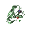
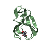
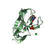
| Entry | Database: PDB / ID: 2a6s | ||||||
|---|---|---|---|---|---|---|---|
| Title | Crystal structure of YoeB under isopropanol condition | ||||||
 Components Components | Toxin yoeB | ||||||
 Keywords Keywords | TOXIN / YoeB / YefM / antitoxin / addiction modules / RNase / inhibitor | ||||||
| Function / homology |  Function and homology information Function and homology informationtoxin-antitoxin complex / global gene silencing by mRNA cleavage / single-species biofilm formation / regulation of growth / mRNA catabolic process / ribosomal small subunit binding / RNA endonuclease activity / negative regulation of translational initiation / response to heat / RNA endonuclease activity producing 3'-phosphomonoesters, hydrolytic mechanism ...toxin-antitoxin complex / global gene silencing by mRNA cleavage / single-species biofilm formation / regulation of growth / mRNA catabolic process / ribosomal small subunit binding / RNA endonuclease activity / negative regulation of translational initiation / response to heat / RNA endonuclease activity producing 3'-phosphomonoesters, hydrolytic mechanism / endonuclease activity / Hydrolases; Acting on ester bonds / regulation of DNA-templated transcription / protein homodimerization activity / RNA binding Similarity search - Function | ||||||
| Biological species |  | ||||||
| Method |  X-RAY DIFFRACTION / SYNCHROTRON / MOLECULAR REPLACEMENT / Resolution: 1.77 Å X-RAY DIFFRACTION / SYNCHROTRON / MOLECULAR REPLACEMENT / Resolution: 1.77 Å | ||||||
 Authors Authors | Kamada, K. / Hanaoka, F. | ||||||
 Citation Citation | Journal: Mol.Cell / Year: 2005 Title: Conformational Change in the Catalytic Site of the Ribonuclease YoeB Toxin by YefM Antitoxin Authors: Kamada, K. / Hanaoka, F. | ||||||
| History |
|
- Structure visualization
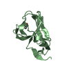
| Structure viewer | Molecule: MolmilJmol/JSmol |
|---|
- Downloads & links
-Download
| PDBx/mmCIF format | 2a6s.cif.gz | 89.7 KB | Display | PDBx/mmCIF format |
|---|---|---|---|---|
| PDB format | pdb2a6s.ent.gz | 68.1 KB | Display | PDB format |
| PDBx/mmJSON format | 2a6s.json.gz | Tree view | PDBx/mmJSON format | |
| Others |  Other downloads Other downloads |
-Validation report
| Arichive directory | https://data.pdbj.org/pub/pdb/validation_reports/a6/2a6sftp://data.pdbj.org/pub/pdb/validation_reports/a6/2a6s | HTTPS FTP |
|---|
-Related structure data
| Related structure data |  2a6qSC  2a6rC S: Starting model for refinement C: citing same article ( |
|---|---|
| Similar structure data |


-Links
 PDBj
PDBj
- Assembly
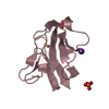
| Deposited unit | 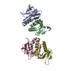
| ||||||||
|---|---|---|---|---|---|---|---|---|---|
| 1 | 
| ||||||||
| 2 | 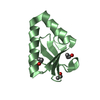
| ||||||||
| 3 | 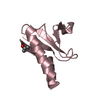
| ||||||||
| 4 | 
| ||||||||
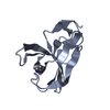
| Unit cell |
|
-Components
| #1: Protein | Mass: 10233.658 Da / Num. of mol.: 4 Source method: isolated from a genetically manipulated source Source: (gene. exp.) #2: Chemical | ChemComp-IPA /   Mass: 60.095 Da / Num. of mol.: 6 / Source method: obtained synthetically / Formula: C3H8O Mass: 60.095 Da / Num. of mol.: 6 / Source method: obtained synthetically / Formula: C3H8O#3: Water | ChemComp-HOH / |  Mass: 18.015 Da / Num. of mol.: 314 / Source method: isolated from a natural source / Formula: H2O Mass: 18.015 Da / Num. of mol.: 314 / Source method: isolated from a natural source / Formula: H2O |
|---|
-Experimental details
-Experiment
| Experiment | Method: X-RAY DIFFRACTION / Number of used crystals: 1 |
|---|
- Sample preparation
| Crystal | Density Matthews: 2.1 Å3/Da / Density % sol: 41.3 % |
|---|---|
| Crystal grow | Temperature: 277.2 K / Method: vapor diffusion, hanging drop / pH: 8.5 Details: Tris-HCl, CH3COONH4, isopropanol, pH 8.5, VAPOR DIFFUSION, HANGING DROP, temperature 277.2K |
-Data collection
| Diffraction | Mean temperature: 100 K |
|---|---|
| Diffraction source | Source: SYNCHROTRON / Site: Photon Factory  / Beamline: BL-5A / Wavelength: 1 Å / Beamline: BL-5A / Wavelength: 1 Å |
| Detector | Type: ADSC QUANTUM 315 / Detector: CCD / Date: May 28, 2004 / Details: Collimating and Focusing mirrors |
| Radiation | Monochromator: Numerical link type Si(111) double crystal monochromator Protocol: SINGLE WAVELENGTH / Monochromatic (M) / Laue (L): M / Scattering type: x-ray |
| Radiation wavelength | Wavelength: 1 Å / Relative weight: 1 |
| Reflection | Resolution: 1.77→48.8 Å / Num. all: 33197 / Num. obs: 33168 / % possible obs: 99.7 % / Observed criterion σ(F): 0 / Observed criterion σ(I): 0 / Redundancy: 7.4 % / Biso Wilson estimate: 14.8 Å2 / Χ2: 1.339 / Net I/σ(I): 42.01 |
| Reflection shell | Resolution: 1.77→1.83 Å / % possible obs: 97.5 % / Redundancy: 6.8 % / Num. measured obs: 3248 / Num. unique all: 3248 / Χ2: 0.954 / % possible all: 97.5 |
-Phasing
| Phasing MR | Cor.coef. Fo:Fc: 0.489 / Packing: 0.624
|
|---|
- Processing
| Software |
| ||||||||||||||||||||||||||||||||||||||||||||||||||||||||||||||||||||||||||||||||||||||||||
|---|---|---|---|---|---|---|---|---|---|---|---|---|---|---|---|---|---|---|---|---|---|---|---|---|---|---|---|---|---|---|---|---|---|---|---|---|---|---|---|---|---|---|---|---|---|---|---|---|---|---|---|---|---|---|---|---|---|---|---|---|---|---|---|---|---|---|---|---|---|---|---|---|---|---|---|---|---|---|---|---|---|---|---|---|---|---|---|---|---|---|---|
| Refinement | Method to determine structure: MOLECULAR REPLACEMENT Starting model: extracted from YoeB structure from 2A6Q Resolution: 1.77→19.96 Å / Rfactor Rfree error: 0.006 / Occupancy max: 1 / Occupancy min: 0 / Cross valid method: THROUGHOUT / σ(F): 2 / Stereochemistry target values: Engh & Huber
| ||||||||||||||||||||||||||||||||||||||||||||||||||||||||||||||||||||||||||||||||||||||||||
| Solvent computation | Solvent model: CNS bulk solvent model used / Bsol: 55.633 Å2 / ksol: 0.446303 e/Å3 | ||||||||||||||||||||||||||||||||||||||||||||||||||||||||||||||||||||||||||||||||||||||||||
| Displacement parameters | Biso max: 55.56 Å2 / Biso mean: 17.15 Å2 / Biso min: 4.72 Å2
| ||||||||||||||||||||||||||||||||||||||||||||||||||||||||||||||||||||||||||||||||||||||||||
| Refine analyze |
| ||||||||||||||||||||||||||||||||||||||||||||||||||||||||||||||||||||||||||||||||||||||||||
| Refinement step | Cycle: LAST / Resolution: 1.77→19.96 Å
| ||||||||||||||||||||||||||||||||||||||||||||||||||||||||||||||||||||||||||||||||||||||||||
| Refine LS restraints |
| ||||||||||||||||||||||||||||||||||||||||||||||||||||||||||||||||||||||||||||||||||||||||||
| LS refinement shell | Refine-ID: X-RAY DIFFRACTION / Total num. of bins used: 8
| ||||||||||||||||||||||||||||||||||||||||||||||||||||||||||||||||||||||||||||||||||||||||||
| Xplor file |
|
