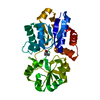
 Movie
Movie Controller


+ Open data

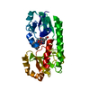
- Basic information
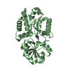
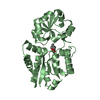
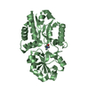
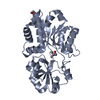
| Entry | Database: PDB / ID: 6eyq | ||||||
|---|---|---|---|---|---|---|---|
| Title | Crystal structure of a mutated OpuBC in complex with choline | ||||||
 Components Components | Choline-binding protein | ||||||
 Keywords Keywords | TRANSPORT PROTEIN / substrate binding protein | ||||||
| Function / homology |  Function and homology information Function and homology informationcellular response to sulfate starvation / amino acid transport / transmembrane transporter activity / ATP-binding cassette (ABC) transporter complex Similarity search - Function | ||||||
| Biological species |  | ||||||
| Method |  X-RAY DIFFRACTION / SYNCHROTRON / MOLECULAR REPLACEMENT / Resolution: 1.5 Å X-RAY DIFFRACTION / SYNCHROTRON / MOLECULAR REPLACEMENT / Resolution: 1.5 Å | ||||||
 Authors Authors | Peherstorfer, S. / Teichmann, L. / Smits, S.H. / Schmitt, L. / Bremer, E. | ||||||
 Citation Citation | Journal: To Be Published Title: Crystal structure of a mutated OpuBC in complex with choline Authors: Peherstorfer, S. / Teichmann, L. / Smits, S.H. / Schmitt, L. / Bremer, E. | ||||||
| History |
|
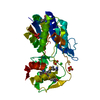
- Structure visualization
| Structure viewer | Molecule: MolmilJmol/JSmol |
|---|
- Downloads & links
-Download
| PDBx/mmCIF format | 6eyq.cif.gz | 233.4 KB | Display | PDBx/mmCIF format |
|---|---|---|---|---|
| PDB format | pdb6eyq.ent.gz | 188.9 KB | Display | PDB format |
| PDBx/mmJSON format | 6eyq.json.gz | Tree view | PDBx/mmJSON format | |
| Others |  Other downloads Other downloads |
-Validation report
| Arichive directory | https://data.pdbj.org/pub/pdb/validation_reports/ey/6eyqftp://data.pdbj.org/pub/pdb/validation_reports/ey/6eyq | HTTPS FTP |
|---|
-Related structure data
| Related structure data |  3r6uS S: Starting model for refinement |
|---|---|
| Similar structure data |

-Links
 PDBj
PDBj


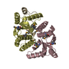
- Assembly
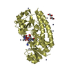
| Deposited unit | 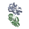
| ||||||||
|---|---|---|---|---|---|---|---|---|---|
| 1 | 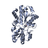
| ||||||||
| 2 | 
| ||||||||
| Unit cell |
|
-Components
| #1: Protein | Mass: 34430.543 Da / Num. of mol.: 2 Source method: isolated from a genetically manipulated source Details: WE mutated D74 to Threonine Source: (gene. exp.) Strain: 168 / Gene: opuBC, proX, BSU33710 / Production host: #2: Chemical | 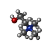  Mass: 104.171 Da / Num. of mol.: 2 / Source method: obtained synthetically / Formula: C5H14NO Mass: 104.171 Da / Num. of mol.: 2 / Source method: obtained synthetically / Formula: C5H14NO#3: Water | ChemComp-HOH / |  Mass: 18.015 Da / Num. of mol.: 713 / Source method: isolated from a natural source / Formula: H2O Mass: 18.015 Da / Num. of mol.: 713 / Source method: isolated from a natural source / Formula: H2O |
|---|
-Experimental details
-Experiment
| Experiment | Method: X-RAY DIFFRACTION / Number of used crystals: 1 |
|---|
- Sample preparation
| Crystal grow | Temperature: 273 K / Method: batch mode / pH: 5 / Details: PEG3350 phosphatte buffer / PH range: 4-6 |
|---|
-Data collection
| Diffraction | Mean temperature: 100 K |
|---|---|
| Diffraction source | Source: SYNCHROTRON / Site: ESRF  / Beamline: ID23-1 / Wavelength: 0.973 Å / Beamline: ID23-1 / Wavelength: 0.973 Å |
| Detector | Type: MAR CCD 130 mm / Detector: CCD / Date: Nov 18, 2015 |
| Radiation | Protocol: SINGLE WAVELENGTH / Monochromatic (M) / Laue (L): M / Scattering type: x-ray |
| Radiation wavelength | Wavelength: 0.973 Å / Relative weight: 1 |
| Reflection | Resolution: 1.5→42.1 Å / Num. obs: 77957 / % possible obs: 99.3 % / Redundancy: 6.5 % / Rsym value: 0.048 / Net I/σ(I): 25.6 |
| Reflection shell | Resolution: 1.5→1.55 Å / Redundancy: 5.4 % / Mean I/σ(I) obs: 12 / Num. unique obs: 7783 / Rsym value: 0.14 / % possible all: 99.3 |
- Processing
| Software |
| ||||||||||||||||||||||||||||||||||||||||||
|---|---|---|---|---|---|---|---|---|---|---|---|---|---|---|---|---|---|---|---|---|---|---|---|---|---|---|---|---|---|---|---|---|---|---|---|---|---|---|---|---|---|---|---|
| Refinement | Method to determine structure: MOLECULAR REPLACEMENT Starting model: 3R6U Resolution: 1.5→42.1 Å / SU ML: 0.09 / Cross valid method: FREE R-VALUE / σ(F): 1.36 / Phase error: 17.43
| ||||||||||||||||||||||||||||||||||||||||||
| Solvent computation | Shrinkage radii: 0.9 Å / VDW probe radii: 1.11 Å | ||||||||||||||||||||||||||||||||||||||||||
| Refinement step | Cycle: LAST / Resolution: 1.5→42.1 Å
| ||||||||||||||||||||||||||||||||||||||||||
| Refine LS restraints |
| ||||||||||||||||||||||||||||||||||||||||||
| LS refinement shell |
|
