 Movie
Movie Controller

[English] 日本語
 Yorodumi
- PDB-6exh: Crystal structure of the complex Fe(II)/alpha-ketoglutarate depen... -
+ Open data

- Basic information
| Entry | Database: PDB / ID: 6exh | ||||||
|---|---|---|---|---|---|---|---|
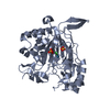
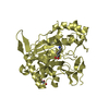
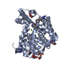
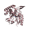
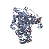
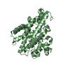
| Title | Crystal structure of the complex Fe(II)/alpha-ketoglutarate dependent dioxygenase KDO5 with Fe(II)/succinate/(4R)-4-hydroxy-L-lysine | ||||||
 Components Components | L-lysine 4-hydroxylase | ||||||
 Keywords Keywords | OXIDOREDUCTASE / Fe(II)/alpha-ketoglutarate / dioxygenases / enzyme / FeII alphaKG form / oxydoreductase | ||||||
| Function / homology | Taurine dioxygenase TauD-like superfamily / 2-oxoglutarate-dependent dioxygenase activity / Oxidoreductases; Acting on paired donors, with incorporation or reduction of molecular oxygen; With 2-oxoglutarate as one donor, and incorporation of one atom of oxygen into each donor / metal ion binding / : / 4-HYDROXY-LYSINE / SUCCINIC ACID / L-lysine 4-hydroxylase Function and homology information Function and homology information | ||||||
| Biological species |  Flavobacterium sp. (bacteria) Flavobacterium sp. (bacteria) | ||||||
| Method |  X-RAY DIFFRACTION / SYNCHROTRON / MOLECULAR REPLACEMENT / Resolution: 2.6 Å X-RAY DIFFRACTION / SYNCHROTRON / MOLECULAR REPLACEMENT / Resolution: 2.6 Å | ||||||
 Authors Authors | Isabet, T. / Stura, E. / Legrand, P. / Zaparucha, A. / Bastard, K. | ||||||
 Citation Citation | Journal: Sci Rep / Year: 2018 Title: Structural Studies based on two Lysine Dioxygenases with Distinct Regioselectivity Brings Insights Into Enzyme Specificity within the Clavaminate Synthase-Like Family. Authors: Bastard, K. / Isabet, T. / Stura, E.A. / Legrand, P. / Zaparucha, A. | ||||||
| History |
|
- Structure visualization
| Structure viewer | Molecule: MolmilJmol/JSmol |
|---|
- Downloads & links
-Download
| PDBx/mmCIF format | 6exh.cif.gz | 580.3 KB | Display | PDBx/mmCIF format |
|---|---|---|---|---|
| PDB format | pdb6exh.ent.gz | 482.5 KB | Display | PDB format |
| PDBx/mmJSON format | 6exh.json.gz | Tree view | PDBx/mmJSON format | |
| Others |  Other downloads Other downloads |
-Validation report
| Arichive directory | https://data.pdbj.org/pub/pdb/validation_reports/ex/6exhftp://data.pdbj.org/pub/pdb/validation_reports/ex/6exh | HTTPS FTP |
|---|
-Related structure data
| Related structure data | 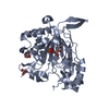 6euoC 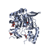 6eurC 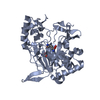 6exfC  6f2aC 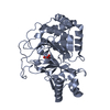 6f2bC  6f2eC 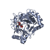 6f6jC  6f9pC C: citing same article ( |
|---|---|
| Similar structure data |
-Links
 PDBj
PDBj- Assembly
| Deposited unit | 
| ||||||||
|---|---|---|---|---|---|---|---|---|---|
| 1 | 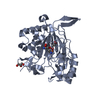
| ||||||||
| 2 | 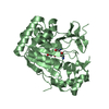
| ||||||||
| 3 | 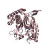
| ||||||||
| 4 | 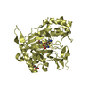
| ||||||||
| Unit cell |
|
-Components
-Protein , 1 types, 4 molecules ABCD
| #1: Protein | Mass: 41817.129 Da / Num. of mol.: 4 Source method: isolated from a genetically manipulated source Source: (gene. exp.) Flavobacterium sp. (bacteria) / Gene: PMI10_03368 / Production host: References: UniProt: J3BZS6, Oxidoreductases; Acting on paired donors, with incorporation or reduction of molecular oxygen; With 2-oxoglutarate as one donor, and incorporation of one atom of oxygen into each donor |
|---|
-Non-polymers , 5 types, 448 molecules 


| #2: Chemical | ChemComp-FE /  Mass: 55.845 Da / Num. of mol.: 4 / Source method: obtained synthetically / Formula: Fe Mass: 55.845 Da / Num. of mol.: 4 / Source method: obtained synthetically / Formula: Fe#3: Chemical | ChemComp-LYO /  Type: L-peptide linking / Mass: 162.187 Da / Num. of mol.: 4 / Source method: obtained synthetically / Formula: C6H14N2O3 Type: L-peptide linking / Mass: 162.187 Da / Num. of mol.: 4 / Source method: obtained synthetically / Formula: C6H14N2O3#4: Chemical |  Mass: 118.088 Da / Num. of mol.: 3 / Source method: obtained synthetically / Formula: C4H6O4 Mass: 118.088 Da / Num. of mol.: 3 / Source method: obtained synthetically / Formula: C4H6O4#5: Chemical |  Mass: 92.094 Da / Num. of mol.: 3 / Source method: obtained synthetically / Formula: C3H8O3 Mass: 92.094 Da / Num. of mol.: 3 / Source method: obtained synthetically / Formula: C3H8O3#6: Water | ChemComp-HOH / | Mass: 18.015 Da / Num. of mol.: 434 / Source method: isolated from a natural source / Formula: H2O |
|---|
-Experimental details
-Experiment
| Experiment | Method: X-RAY DIFFRACTION / Number of used crystals: 1 |
|---|
- Sample preparation
| Crystal | Density Matthews: 2.25 Å3/Da / Density % sol: 45.4 % |
|---|---|
| Crystal grow | Temperature: 293 K / Method: vapor diffusion, sitting drop Details: 25% PEG4000, 0.2M Imidazole malate pH7, 0.15M Li2SO4 |
-Data collection
| Diffraction | Mean temperature: 100 K |
|---|---|
| Diffraction source | Source: SYNCHROTRON / Site: SOLEIL  / Beamline: PROXIMA 2 / Wavelength: 0.9801 Å / Beamline: PROXIMA 2 / Wavelength: 0.9801 Å |
| Detector | Type: DECTRIS EIGER X 9M / Detector: PIXEL / Date: Jul 8, 2016 |
| Radiation | Protocol: SINGLE WAVELENGTH / Monochromatic (M) / Laue (L): M / Scattering type: x-ray |
| Radiation wavelength | Wavelength: 0.9801 Å / Relative weight: 1 |
| Reflection | Resolution: 2.6→63.57 Å / Num. obs: 47269 / % possible obs: 99.8 % / Redundancy: 13.7 % / CC1/2: 0.999 / Rrim(I) all: 0.165 / Net I/σ(I): 13.86 |
| Reflection shell | Resolution: 2.6→2.76 Å / Mean I/σ(I) obs: 1.4 / Num. unique obs: 7484 / CC1/2: 0.84 / Rrim(I) all: 1.56 / % possible all: 99 |
- Processing
| Software |
| |||||||||||||||||||||||||||||||||||||||||||||||||||||||||||||||||||||||||||||||||||||||||||||||||||||||||||||||||||||||||||||
|---|---|---|---|---|---|---|---|---|---|---|---|---|---|---|---|---|---|---|---|---|---|---|---|---|---|---|---|---|---|---|---|---|---|---|---|---|---|---|---|---|---|---|---|---|---|---|---|---|---|---|---|---|---|---|---|---|---|---|---|---|---|---|---|---|---|---|---|---|---|---|---|---|---|---|---|---|---|---|---|---|---|---|---|---|---|---|---|---|---|---|---|---|---|---|---|---|---|---|---|---|---|---|---|---|---|---|---|---|---|---|---|---|---|---|---|---|---|---|---|---|---|---|---|---|---|---|
| Refinement | Method to determine structure: MOLECULAR REPLACEMENT / Resolution: 2.6→38.07 Å / Cor.coef. Fo:Fc: 0.94 / Cor.coef. Fo:Fc free: 0.921 / SU R Cruickshank DPI: 1.812 / Cross valid method: THROUGHOUT / σ(F): 0 / SU Rfree Blow DPI: 0.289 / SU Rfree Cruickshank DPI: 0.291
| |||||||||||||||||||||||||||||||||||||||||||||||||||||||||||||||||||||||||||||||||||||||||||||||||||||||||||||||||||||||||||||
| Displacement parameters | Biso mean: 79.54 Å2
| |||||||||||||||||||||||||||||||||||||||||||||||||||||||||||||||||||||||||||||||||||||||||||||||||||||||||||||||||||||||||||||
| Refine analyze | Luzzati coordinate error obs: 0.32 Å | |||||||||||||||||||||||||||||||||||||||||||||||||||||||||||||||||||||||||||||||||||||||||||||||||||||||||||||||||||||||||||||
| Refinement step | Cycle: 1 / Resolution: 2.6→38.07 Å
| |||||||||||||||||||||||||||||||||||||||||||||||||||||||||||||||||||||||||||||||||||||||||||||||||||||||||||||||||||||||||||||
| Refine LS restraints |
| |||||||||||||||||||||||||||||||||||||||||||||||||||||||||||||||||||||||||||||||||||||||||||||||||||||||||||||||||||||||||||||
| LS refinement shell | Resolution: 2.6→2.67 Å / Total num. of bins used: 20
| |||||||||||||||||||||||||||||||||||||||||||||||||||||||||||||||||||||||||||||||||||||||||||||||||||||||||||||||||||||||||||||
| Refinement TLS params. | Method: refined / Refine-ID: X-RAY DIFFRACTION
| |||||||||||||||||||||||||||||||||||||||||||||||||||||||||||||||||||||||||||||||||||||||||||||||||||||||||||||||||||||||||||||
| Refinement TLS group |
|
