 Movie
Movie Controller


+ Open data

- Basic information
| Entry | Database: PDB / ID: 6et5 | |||||||||||||||||||||||||||||||||||||||||||||||||||||||||||||||||||||||||||||||||||||||||||||||||||||||||||||||||||||
|---|---|---|---|---|---|---|---|---|---|---|---|---|---|---|---|---|---|---|---|---|---|---|---|---|---|---|---|---|---|---|---|---|---|---|---|---|---|---|---|---|---|---|---|---|---|---|---|---|---|---|---|---|---|---|---|---|---|---|---|---|---|---|---|---|---|---|---|---|---|---|---|---|---|---|---|---|---|---|---|---|---|---|---|---|---|---|---|---|---|---|---|---|---|---|---|---|---|---|---|---|---|---|---|---|---|---|---|---|---|---|---|---|---|---|---|---|---|---|
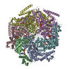
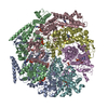
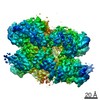
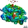
| Title | Reaction centre light harvesting complex 1 from Blc. virids | |||||||||||||||||||||||||||||||||||||||||||||||||||||||||||||||||||||||||||||||||||||||||||||||||||||||||||||||||||||
 Components Components |
| |||||||||||||||||||||||||||||||||||||||||||||||||||||||||||||||||||||||||||||||||||||||||||||||||||||||||||||||||||||
 Keywords Keywords | PHOTOSYNTHESIS / Reaction centre light harvesting complex 1 Blc. viridis Cryo-EM RC-LH1 Photosynthesis | |||||||||||||||||||||||||||||||||||||||||||||||||||||||||||||||||||||||||||||||||||||||||||||||||||||||||||||||||||||
| Function / homology |  Function and homology information Function and homology informationlight-harvesting complex / organelle inner membrane / plasma membrane-derived chromatophore membrane / plasma membrane light-harvesting complex / bacteriochlorophyll binding / photosynthetic electron transport in photosystem II / photosynthesis, light reaction / photosynthesis / electron transfer activity / iron ion binding ...light-harvesting complex / organelle inner membrane / plasma membrane-derived chromatophore membrane / plasma membrane light-harvesting complex / bacteriochlorophyll binding / photosynthetic electron transport in photosystem II / photosynthesis, light reaction / photosynthesis / electron transfer activity / iron ion binding / heme binding / metal ion binding / plasma membrane Similarity search - Function | |||||||||||||||||||||||||||||||||||||||||||||||||||||||||||||||||||||||||||||||||||||||||||||||||||||||||||||||||||||
| Biological species |  Blastochloris viridis (bacteria) Blastochloris viridis (bacteria) | |||||||||||||||||||||||||||||||||||||||||||||||||||||||||||||||||||||||||||||||||||||||||||||||||||||||||||||||||||||
| Method | ELECTRON MICROSCOPY / single particle reconstruction / cryo EM / Resolution: 2.87 Å | |||||||||||||||||||||||||||||||||||||||||||||||||||||||||||||||||||||||||||||||||||||||||||||||||||||||||||||||||||||
 Authors Authors | Qian, P. / Siebert, C.A. / Canniffe, D.P. / Wang, P. / Hunter, C.N. | |||||||||||||||||||||||||||||||||||||||||||||||||||||||||||||||||||||||||||||||||||||||||||||||||||||||||||||||||||||
| Funding support | 1items
| |||||||||||||||||||||||||||||||||||||||||||||||||||||||||||||||||||||||||||||||||||||||||||||||||||||||||||||||||||||
 Citation Citation | Journal: Nature / Year: 2018 Title: Cryo-EM structure of the Blastochloris viridis LH1-RC complex at 2.9 Å. Authors: Pu Qian / C Alistair Siebert / Peiyi Wang / Daniel P Canniffe / C Neil Hunter /  Abstract: The light-harvesting 1-reaction centre (LH1-RC) complex is a key functional component of bacterial photosynthesis. Here we present a 2.9 Å resolution cryo-electron microscopy structure of the ...The light-harvesting 1-reaction centre (LH1-RC) complex is a key functional component of bacterial photosynthesis. Here we present a 2.9 Å resolution cryo-electron microscopy structure of the bacteriochlorophyll b-based LH1-RC complex from Blastochloris viridis that reveals the structural basis for absorption of infrared light and the molecular mechanism of quinone migration across the LH1 complex. The triple-ring LH1 complex comprises a circular array of 17 β-polypeptides sandwiched between 17 α- and 16 γ-polypeptides. Tight packing of the γ-apoproteins between β-polypeptides collectively interlocks and stabilizes the LH1 structure; this, together with the short Mg-Mg distances of bacteriochlorophyll b pairs, contributes to the large redshift of bacteriochlorophyll b absorption. The 'missing' 17th γ-polypeptide creates a pore in the LH1 ring, and an adjacent binding pocket provides a folding template for a quinone, Q , which adopts a compact, export-ready conformation before passage through the pore and eventual diffusion to the cytochrome bc complex. | |||||||||||||||||||||||||||||||||||||||||||||||||||||||||||||||||||||||||||||||||||||||||||||||||||||||||||||||||||||
| History |
|
- Structure visualization
| Movie |
Movie viewer |
|---|---|
| Structure viewer | Molecule: MolmilJmol/JSmol |
- Downloads & links
-Download
| PDBx/mmCIF format | 6et5.cif.gz | 805 KB | Display | PDBx/mmCIF format |
|---|---|---|---|---|
| PDB format | pdb6et5.ent.gz | 669.8 KB | Display | PDB format |
| PDBx/mmJSON format | 6et5.json.gz | Tree view | PDBx/mmJSON format | |
| Others |  Other downloads Other downloads |
-Validation report
| Arichive directory | https://data.pdbj.org/pub/pdb/validation_reports/et/6et5ftp://data.pdbj.org/pub/pdb/validation_reports/et/6et5 | HTTPS FTP |
|---|
-Related structure data
| Related structure data | 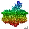 3951MC M: map data used to model this data C: citing same article ( |
|---|---|
| Similar structure data |


-Links
 PDBj
PDBj


- Assembly
| Deposited unit | 
|
|---|---|
| 1 |
|
-Components
-Protein , 1 types, 1 molecules C
| #1: Protein | Mass: 37179.465 Da / Num. of mol.: 1 / Source method: isolated from a natural source / Details: reaction centre cytochrome subunit / Source: (natural) Blastochloris viridis (bacteria) / References: UniProt: P07173 |
|---|
-Reaction center protein ... , 3 types, 3 molecules LMH
| #2: Protein | Mass: 30600.299 Da / Num. of mol.: 1 / Source method: isolated from a natural source / Details: reaction centre L subunit / Source: (natural) Blastochloris viridis (bacteria) / References: UniProt: P06009 |
|---|---|
| #3: Protein | Mass: 35932.188 Da / Num. of mol.: 1 / Source method: isolated from a natural source / Details: Teaction centre M subunit / Source: (natural) Blastochloris viridis (bacteria) / References: UniProt: P06010 |
| #4: Protein | Mass: 28557.453 Da / Num. of mol.: 1 / Source method: isolated from a natural source / Details: Reaction centre H subunit / Source: (natural) Blastochloris viridis (bacteria) / References: UniProt: P06008 |
-Light-harvesting protein B-1015 ... , 3 types, 50 molecules zFKPSVYbehknqtw361GNQTWZcfilor...
| #5: Protein | Mass: 6984.262 Da / Num. of mol.: 17 / Source method: isolated from a natural source / Details: Light harvesting complex 1 alpha subunit / Source: (natural) Blastochloris viridis (bacteria) / References: UniProt: P04123#6: Protein | Mass: 6274.327 Da / Num. of mol.: 17 / Source method: isolated from a natural source / Details: Light harvesting complex 1 beta subunit / Source: (natural) Blastochloris viridis (bacteria) / References: UniProt: P04124#7: Protein/peptide | Mass: 2799.292 Da / Num. of mol.: 16 / Source method: isolated from a natural source / Details: Light harvesting complex 1 gamma subunit / Source: (natural) Blastochloris viridis (bacteria) / References: UniProt: P04126 |
|---|
-Non-polymers , 10 types, 75 molecules 


| #8: Chemical | ChemComp-HEC /  Mass: 618.503 Da / Num. of mol.: 4 / Source method: obtained synthetically / Formula: C34H34FeN4O4 Mass: 618.503 Da / Num. of mol.: 4 / Source method: obtained synthetically / Formula: C34H34FeN4O4#9: Chemical | ChemComp-BCB /  Mass: 909.488 Da / Num. of mol.: 38 / Source method: obtained synthetically / Formula: C55H72MgN4O6 Mass: 909.488 Da / Num. of mol.: 38 / Source method: obtained synthetically / Formula: C55H72MgN4O6#10: Chemical |  Mass: 887.199 Da / Num. of mol.: 2 / Source method: obtained synthetically / Formula: C55H74N4O6 Mass: 887.199 Da / Num. of mol.: 2 / Source method: obtained synthetically / Formula: C55H74N4O6#11: Chemical |  Mass: 795.226 Da / Num. of mol.: 2 / Source method: obtained synthetically / Formula: C54H82O4 Mass: 795.226 Da / Num. of mol.: 2 / Source method: obtained synthetically / Formula: C54H82O4#12: Chemical | ChemComp-FE / |  Mass: 55.845 Da / Num. of mol.: 1 / Source method: obtained synthetically / Formula: Fe Mass: 55.845 Da / Num. of mol.: 1 / Source method: obtained synthetically / Formula: Fe#13: Chemical | ChemComp-SO4 /  Mass: 96.063 Da / Num. of mol.: 7 / Source method: obtained synthetically / Formula: SO4 Mass: 96.063 Da / Num. of mol.: 7 / Source method: obtained synthetically / Formula: SO4#14: Chemical |  Mass: 229.402 Da / Num. of mol.: 2 / Source method: obtained synthetically / Formula: C14H31NO / Comment: LDAO, detergent*YM Mass: 229.402 Da / Num. of mol.: 2 / Source method: obtained synthetically / Formula: C14H31NO / Comment: LDAO, detergent*YM#15: Chemical | ChemComp-MQ9 / |  Mass: 785.233 Da / Num. of mol.: 1 / Source method: obtained synthetically / Formula: C56H80O2 Mass: 785.233 Da / Num. of mol.: 1 / Source method: obtained synthetically / Formula: C56H80O2#16: Chemical | ChemComp-NS5 / |  Mass: 540.904 Da / Num. of mol.: 1 / Source method: obtained synthetically / Formula: C40H60 Mass: 540.904 Da / Num. of mol.: 1 / Source method: obtained synthetically / Formula: C40H60#17: Chemical | ChemComp-NS0 /  Mass: 540.904 Da / Num. of mol.: 17 / Source method: obtained synthetically / Formula: C40H60 Mass: 540.904 Da / Num. of mol.: 17 / Source method: obtained synthetically / Formula: C40H60 |
|---|
-Details
| Has protein modification | Y |
|---|
-Experimental details
-Experiment
| Experiment | Method: ELECTRON MICROSCOPY |
|---|---|
| EM experiment | Aggregation state: CELL / 3D reconstruction method: single particle reconstruction |
- Sample preparation
| Component | Name: Reaction centre-Light harvesting complex 1 (RC-LH1) / Type: COMPLEX Details: The protein was isolated from a photosynthetic bacterium Blc. viridis. Entity ID: #1-#7 / Source: NATURAL |
|---|---|
| Molecular weight | Value: 0.414 MDa / Experimental value: YES |
| Source (natural) | Organism: Blastochloris viridis (bacteria) |
| Buffer solution | pH: 7.8 / Details: 20 mMol HEPES, pH 7.8, 100 mM NaCl |
| Buffer component | Conc.: 20 mMol / Name: HEPES |
| Specimen | Conc.: 4 mg/ml / Embedding applied: NO / Shadowing applied: NO / Staining applied: NO / Vitrification applied: YES Details: Protein was solubilised by the use detergent of beta-DDM. The protein particle therefore contains a detergent belt in its hydrophobic region. |
| Specimen support | Grid material: COPPER / Grid mesh size: 300 divisions/in. / Grid type: Quantifoil R1.2/1.3 |
| Vitrification | Instrument: LEICA EM GP / Cryogen name: ETHANE / Temp: 277 K / Humidity: 99 % / Chamber temperature: 278 K Details: Blot for 3.5 seconds. Humidity: 99% Temperature:4C. |
- Electron microscopy imaging
| Experimental equipment |  Model: Titan Krios / Image courtesy: FEI Company |
|---|---|
| Microscopy | Model: FEI TITAN KRIOS |
| Electron gun | Electron source:  FIELD EMISSION GUN / Accelerating voltage: 300 kV / Illumination mode: FLOOD BEAM FIELD EMISSION GUN / Accelerating voltage: 300 kV / Illumination mode: FLOOD BEAM |
| Electron lens | Mode: BRIGHT FIELD / Calibrated magnification: 130000 X / Cs: 2.7 mm |
| Specimen holder | Cryogen: NITROGEN / Specimen holder model: FEI TITAN KRIOS AUTOGRID HOLDER / Temperature (min): 77 K |
| Image recording | Average exposure time: 0.5 sec. / Electron dose: 2.25 e/Å2 / Detector mode: COUNTING / Film or detector model: GATAN K2 SUMMIT (4k x 4k) / Num. of grids imaged: 3 / Num. of real images: 6472 |
| Image scans | Movie frames/image: 20 / Used frames/image: 1-20 |
- Processing
| Software | Name: REFMAC / Version: 5.8.0158 / Classification: refinement | ||||||||||||||||||||||||||||||||||||||||||||||||||||||||||||||||||||||||||||||||||||||||||||||||||||||||||
|---|---|---|---|---|---|---|---|---|---|---|---|---|---|---|---|---|---|---|---|---|---|---|---|---|---|---|---|---|---|---|---|---|---|---|---|---|---|---|---|---|---|---|---|---|---|---|---|---|---|---|---|---|---|---|---|---|---|---|---|---|---|---|---|---|---|---|---|---|---|---|---|---|---|---|---|---|---|---|---|---|---|---|---|---|---|---|---|---|---|---|---|---|---|---|---|---|---|---|---|---|---|---|---|---|---|---|---|
| EM software |
| ||||||||||||||||||||||||||||||||||||||||||||||||||||||||||||||||||||||||||||||||||||||||||||||||||||||||||
| Image processing | Details: Dose fractioned | ||||||||||||||||||||||||||||||||||||||||||||||||||||||||||||||||||||||||||||||||||||||||||||||||||||||||||
| CTF correction | Details: gctf was used for CTF correction / Type: PHASE FLIPPING AND AMPLITUDE CORRECTION | ||||||||||||||||||||||||||||||||||||||||||||||||||||||||||||||||||||||||||||||||||||||||||||||||||||||||||
| Particle selection | Num. of particles selected: 267726 / Details: Protein was checked using negatively stained EM. | ||||||||||||||||||||||||||||||||||||||||||||||||||||||||||||||||||||||||||||||||||||||||||||||||||||||||||
| Symmetry | Point symmetry: C1 (asymmetric) | ||||||||||||||||||||||||||||||||||||||||||||||||||||||||||||||||||||||||||||||||||||||||||||||||||||||||||
| 3D reconstruction | Resolution: 2.87 Å / Resolution method: FSC 0.143 CUT-OFF / Num. of particles: 267726 / Algorithm: FOURIER SPACE / Symmetry type: POINT | ||||||||||||||||||||||||||||||||||||||||||||||||||||||||||||||||||||||||||||||||||||||||||||||||||||||||||
| Atomic model building | Protocol: BACKBONE TRACE / Space: REAL | ||||||||||||||||||||||||||||||||||||||||||||||||||||||||||||||||||||||||||||||||||||||||||||||||||||||||||
| Refinement | Resolution: 2.87→2.87 Å / Cor.coef. Fo:Fc: 0.875 / SU B: 12.39 / SU ML: 0.188 / ESU R: 0.189 Stereochemistry target values: MAXIMUM LIKELIHOOD WITH PHASES
| ||||||||||||||||||||||||||||||||||||||||||||||||||||||||||||||||||||||||||||||||||||||||||||||||||||||||||
| Solvent computation | Solvent model: PARAMETERS FOR MASK CACLULATION | ||||||||||||||||||||||||||||||||||||||||||||||||||||||||||||||||||||||||||||||||||||||||||||||||||||||||||
| Displacement parameters | Biso mean: 153.894 Å2
| ||||||||||||||||||||||||||||||||||||||||||||||||||||||||||||||||||||||||||||||||||||||||||||||||||||||||||
| Refinement step | Cycle: 1 / Total: 31896 | ||||||||||||||||||||||||||||||||||||||||||||||||||||||||||||||||||||||||||||||||||||||||||||||||||||||||||
| Refine LS restraints |
|