- EMDB-3951: Reaction centre light harvesting complex 1 from Blc. viridis -
+
Open data
ID or keywords:
Loading...
-
Basic information
Entry
Database: EMDB / ID: EMD-3951
Title
Reaction centre light harvesting complex 1 from Blc. viridis
Map data
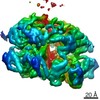
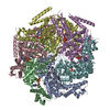
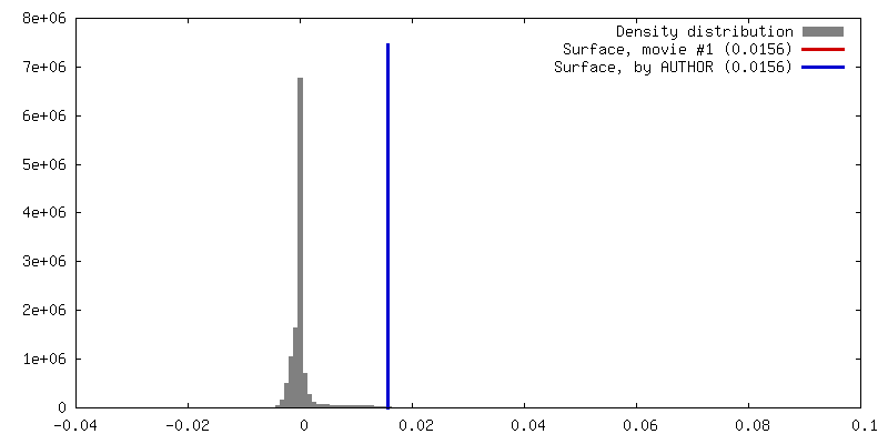
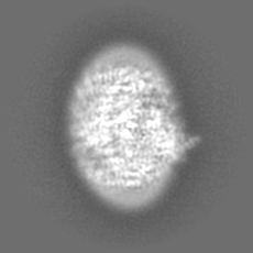
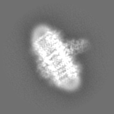
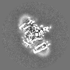
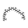
Cryo-EM images of Reaction centre-light harvesting complex 1 (RC-LH1) from Blc. viridis were processed using RELION 2.0. The 3D cryo-EM map was produced after Post-processing to 2.87 Angstrom.
light-harvesting complex / organelle inner membrane / plasma membrane-derived chromatophore membrane / plasma membrane light-harvesting complex / bacteriochlorophyll binding / photosynthetic electron transport in photosystem II / photosynthesis, light reaction / photosynthesis / electron transfer activity / iron ion binding ...light-harvesting complex / organelle inner membrane / plasma membrane-derived chromatophore membrane / plasma membrane light-harvesting complex / bacteriochlorophyll binding / photosynthetic electron transport in photosystem II / photosynthesis, light reaction / photosynthesis / electron transfer activity / iron ion binding / heme binding / metal ion binding / plasma membrane Similarity search - Function
Photosynthetic reaction centre, cytochrome c subunit / Multihaem cytochrome, PRC subunit superfamily / Photosynthetic reaction centre cytochrome C subunit / Antenna complex, beta subunit, conserved site / Antenna complexes beta subunits signature. / Antenna complex, alpha subunit / Antenna complex, alpha subunit conserved site / Antenna complexes alpha subunits signature. / Multiheme cytochrome c family profile. / Antenna complex, alpha/beta subunit ...Photosynthetic reaction centre, cytochrome c subunit / Multihaem cytochrome, PRC subunit superfamily / Photosynthetic reaction centre cytochrome C subunit / Antenna complex, beta subunit, conserved site / Antenna complexes beta subunits signature. / Antenna complex, alpha subunit / Antenna complex, alpha subunit conserved site / Antenna complexes alpha subunits signature. / Multiheme cytochrome c family profile. / Antenna complex, alpha/beta subunit / Light-harvesting protein B beta chain / Antenna complex, beta domain superfamily / Antenna complex alpha/beta subunit / Light-harvesting complex / Photosynthetic reaction centre, H subunit / Bacterial photosynthetic reaction centre, H-chain, C-terminal / Photosynthetic reaction centre, H subunit, N-terminal / PRC-barrel domain / Photosynthetic reaction centre, H subunit, N-terminal domain superfamily / Photosynthetic reaction centre, H-chain N-terminal region / Photosynthetic reaction centre, M subunit / PRC-barrel domain / PRC-barrel-like superfamily / Photosynthetic reaction centre, L subunit / Multiheme cytochrome superfamily / : / Photosynthetic reaction centre, L/M / Photosystem II protein D1/D2 superfamily / Photosynthetic reaction centre protein / Photosynthetic reaction center proteins signature. / Prokaryotic membrane lipoprotein lipid attachment site profile. Similarity search - Domain/homology
Light-harvesting protein B-1015 alpha chain / Light-harvesting protein B-1015 beta chain / Light-harvesting protein B-1015 gamma chain / Reaction center protein H chain / Reaction center protein L chain / Reaction center protein M chain / Photosynthetic reaction center cytochrome c subunit Similarity search - Component
Biological species
Blastochloris viridis (bacteria)
Method
single particle reconstruction / cryo EM / Resolution: 2.87 Å
Journal: Nature / Year: 2018 Title: Cryo-EM structure of the Blastochloris viridis LH1-RC complex at 2.9 Å. Authors: Pu Qian / C Alistair Siebert / Peiyi Wang / Daniel P Canniffe / C Neil Hunter / Abstract: The light-harvesting 1-reaction centre (LH1-RC) complex is a key functional component of bacterial photosynthesis. Here we present a 2.9 Å resolution cryo-electron microscopy structure of the ...The light-harvesting 1-reaction centre (LH1-RC) complex is a key functional component of bacterial photosynthesis. Here we present a 2.9 Å resolution cryo-electron microscopy structure of the bacteriochlorophyll b-based LH1-RC complex from Blastochloris viridis that reveals the structural basis for absorption of infrared light and the molecular mechanism of quinone migration across the LH1 complex. The triple-ring LH1 complex comprises a circular array of 17 β-polypeptides sandwiched between 17 α- and 16 γ-polypeptides. Tight packing of the γ-apoproteins between β-polypeptides collectively interlocks and stabilizes the LH1 structure; this, together with the short Mg-Mg distances of bacteriochlorophyll b pairs, contributes to the large redshift of bacteriochlorophyll b absorption. The 'missing' 17th γ-polypeptide creates a pore in the LH1 ring, and an adjacent binding pocket provides a folding template for a quinone, Q , which adopts a compact, export-ready conformation before passage through the pore and eventual diffusion to the cytochrome bc complex.
History
Deposition
Oct 25, 2017
-
Header (metadata) release
Dec 20, 2017
-
Map release
Apr 11, 2018
-
Update
Oct 1, 2025
-
Current status
Oct 1, 2025
Processing site: PDBe / Status: Released
-
Structure visualization
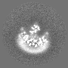
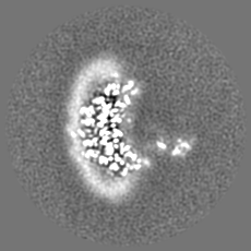
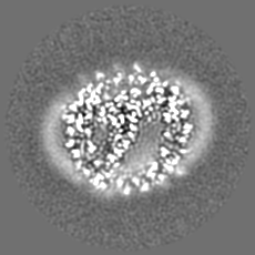
Movie
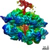
Surface view with section colored by density value
#262 - Oct 2021 Fifty Years of Open Access to PDB Structures similarity (1)
#29 - May 2002 Penicillin-binding Proteins similarity (1)
#95 - Nov 2007 Multidrug Resistance Transporters similarity (1)
-
Map
File
Download / File: emd_3951.map.gz / Format: CCP4 / Size: 46.4 MB / Type: IMAGE STORED AS FLOATING POINT NUMBER (4 BYTES)
Annotation
Cryo-EM images of Reaction centre-light harvesting complex 1 (RC-LH1) from Blc. viridis were processed using RELION 2.0. The 3D cryo-EM map was produced after Post-processing to 2.87 Angstrom.
Model: Quantifoil R1.2/1.3 / Material: COPPER / Mesh: 300 / Support film - Material: CARBON / Support film - topology: CONTINUOUS / Support film - Film thickness: 10 / Pretreatment - Type: GLOW DISCHARGE / Pretreatment - Time: 30 sec. / Pretreatment - Atmosphere: AIR / Pretreatment - Pressure: 0.0002 kPa
Vitrification
Cryogen name: ETHANE / Chamber humidity: 99 % / Chamber temperature: 278 K / Instrument: LEICA EM GP Details: Blot for 3.5 seconds. Humidity: 99% Temperature:4C..
Details
Protein was solubilised by the use detergent of beta-DDM. The protein particle therefore contains a detergent belt in its hydrophobic region.
-
Electron microscopy
Microscope
FEI TITAN KRIOS
Temperature
Min: 77.0 K
Image recording
Film or detector model: GATAN K2 SUMMIT (4k x 4k) / Detector mode: COUNTING / Digitization - Frames/image: 1-20 / Number grids imaged: 3 / Number real images: 6472 / Average exposure time: 0.5 sec. / Average electron dose: 2.25 e/Å2
Electron beam
Acceleration voltage: 300 kV / Electron source: FIELD EMISSION GUN
Electron optics
Calibrated magnification: 130000 / Illumination mode: FLOOD BEAM / Imaging mode: BRIGHT FIELD / Cs: 2.7 mm
In the structure databanks used in Yorodumi, some data are registered as the other names, "COVID-19 virus" and "2019-nCoV". Here are the details of the virus and the list of structure data.
Jan 31, 2019. EMDB accession codes are about to change! (news from PDBe EMDB page)
EMDB accession codes are about to change! (news from PDBe EMDB page)
The allocation of 4 digits for EMDB accession codes will soon come to an end. Whilst these codes will remain in use, new EMDB accession codes will include an additional digit and will expand incrementally as the available range of codes is exhausted. The current 4-digit format prefixed with “EMD-” (i.e. EMD-XXXX) will advance to a 5-digit format (i.e. EMD-XXXXX), and so on. It is currently estimated that the 4-digit codes will be depleted around Spring 2019, at which point the 5-digit format will come into force.
The EM Navigator/Yorodumi systems omit the EMD- prefix.
Related info.:Q: What is EMD? / ID/Accession-code notation in Yorodumi/EM Navigator
Yorodumi is a browser for structure data from EMDB, PDB, SASBDB, etc.
This page is also the successor to EM Navigator detail page, and also detail information page/front-end page for Omokage search.
The word "yorodu" (or yorozu) is an old Japanese word meaning "ten thousand". "mi" (miru) is to see.
Related info.:EMDB / PDB / SASBDB / Comparison of 3 databanks / Yorodumi Search / Aug 31, 2016. New EM Navigator & Yorodumi / Yorodumi Papers / Jmol/JSmol / Function and homology information / Changes in new EM Navigator and Yorodumi
 Movie
Movie Controller


 Open data
 Basic information
 Map data
 Sample
 Keywords
 Function and homology information
 Blastochloris viridis (bacteria)
 Authors
 Citation
 Structure visualization
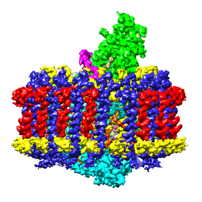
 Downloads & links
 emd_3951.png
 http://ftp.pdbj.org/pub/emdb/structures/EMD-3951


 Z (Sec.)
 Y (Row.)
 X (Col.)


 Sample components


 Processing
 Electron microscopy
 FIELD EMISSION GUN

