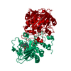
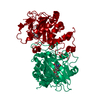
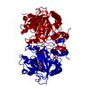
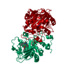
Entry Database : PDB / ID : 6eozTitle Fe(II)/(alpha)ketoglutarate-dependent dioxygenase AsqJ_V72K mutant in complex with cyclopeptin (1b) Iron/alpha-ketoglutarate-dependent dioxygenase asqJ Keywords / / / / / / / Function / homology Function Domain/homology Component
Biological species Emericella nidulans (mold)Method / / / Resolution : 1.55 Å Authors Groll, M. / Braeuer, A. / Kaila, V.R.I. Funding support Organization Grant number Country German Research Foundation SFB1035
Journal : Nat Commun / Year : 2018Title : Catalytic mechanism and molecular engineering of quinolone biosynthesis in dioxygenase AsqJ.Authors : Mader, S.L. / Brauer, A. / Groll, M. / Kaila, V.R.I. History Deposition Oct 10, 2017 Deposition site / Processing site Revision 1.0 Apr 4, 2018 Provider / Type Revision 1.1 Jan 17, 2024 Group Data collection / Database references ... Data collection / Database references / Derived calculations / Refinement description Category chem_comp_atom / chem_comp_bond ... chem_comp_atom / chem_comp_bond / database_2 / pdbx_initial_refinement_model / pdbx_struct_conn_angle / struct_conn Item _database_2.pdbx_DOI / _database_2.pdbx_database_accession ... _database_2.pdbx_DOI / _database_2.pdbx_database_accession / _pdbx_struct_conn_angle.ptnr1_auth_comp_id / _pdbx_struct_conn_angle.ptnr1_auth_seq_id / _pdbx_struct_conn_angle.ptnr1_label_asym_id / _pdbx_struct_conn_angle.ptnr1_label_atom_id / _pdbx_struct_conn_angle.ptnr1_label_comp_id / _pdbx_struct_conn_angle.ptnr3_auth_comp_id / _pdbx_struct_conn_angle.ptnr3_auth_seq_id / _pdbx_struct_conn_angle.ptnr3_label_asym_id / _pdbx_struct_conn_angle.ptnr3_label_atom_id / _pdbx_struct_conn_angle.ptnr3_label_comp_id / _pdbx_struct_conn_angle.value / _struct_conn.pdbx_dist_value / _struct_conn.ptnr2_auth_comp_id / _struct_conn.ptnr2_auth_seq_id / _struct_conn.ptnr2_label_asym_id / _struct_conn.ptnr2_label_atom_id / _struct_conn.ptnr2_label_comp_id
Show all Show less
 Movie
Movie Controller

 Yorodumi
 Open data
 Basic information
 Components
 Keywords
 Function and homology information
 X-RAY DIFFRACTION /
  Authors
 Germany, 1items
  Citation
 Structure visualization
 Downloads & links
 Other downloads
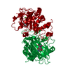


 PDBj
PDBj

 Assembly


 Mass: 58.693 Da / Num. of mol.: 1 / Source method: obtained synthetically / Formula: Ni
 Mass: 146.098 Da / Num. of mol.: 1 / Source method: obtained synthetically / Formula: C5H6O5
 Mass: 280.321 Da / Num. of mol.: 1 / Source method: obtained synthetically / Formula: C17H16N2O2
 Mass: 18.015 Da / Num. of mol.: 301 / Source method: isolated from a natural source / Formula: H2O
 Sample preparation
 / Beamline: X06SA / Wavelength: 1 Å
 Processing