 Movie
Movie Controller

[English] 日本語
 Yorodumi
- PDB-5lk6: Crystal structure of a lipase carboxylesterase from Sulfolobus is... -
+ Open data

- Basic information
| Entry | Database: PDB / ID: 5lk6 | ||||||
|---|---|---|---|---|---|---|---|
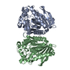
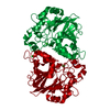
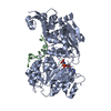
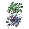
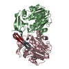
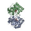
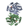
| Title | Crystal structure of a lipase carboxylesterase from Sulfolobus islandicus | ||||||
 Components Components | Alpha/beta hydrolase fold-3 domain protein | ||||||
 Keywords Keywords | HYDROLASE / Esterase / lipase / Sulfolobus / thermostable | ||||||
| Function / homology |  Function and homology information Function and homology information | ||||||
| Biological species |   Sulfolobus islandicus (archaea) Sulfolobus islandicus (archaea) | ||||||
| Method |  X-RAY DIFFRACTION / SYNCHROTRON / MOLECULAR REPLACEMENT / Resolution: 2.6 Å X-RAY DIFFRACTION / SYNCHROTRON / MOLECULAR REPLACEMENT / Resolution: 2.6 Å | ||||||
 Authors Authors | Schwarz-Linnet, T. / Teilum, K. / Olsen, J.G. | ||||||
| Funding support |  Denmark, 1items Denmark, 1items
| ||||||
 Citation Citation | Journal: To Be Published Title: EstA from Sufolobus islandicus is stabilized by mono-methylation of lysine residues Authors: Schwarz-Linnet, T. / Stiefler-Jensen, D. / de Lichetenberg, C. / Olsen, J.G. / Nguyen, T.T.T.N. / Rand, K.D. / Teilum, K. | ||||||
| History |
|
- Structure visualization
| Structure viewer | Molecule: MolmilJmol/JSmol |
|---|
- Downloads & links
-Download
| PDBx/mmCIF format | 5lk6.cif.gz | 248.1 KB | Display | PDBx/mmCIF format |
|---|---|---|---|---|
| PDB format | pdb5lk6.ent.gz | 201.6 KB | Display | PDB format |
| PDBx/mmJSON format | 5lk6.json.gz | Tree view | PDBx/mmJSON format | |
| Others |  Other downloads Other downloads |
-Validation report
| Arichive directory | https://data.pdbj.org/pub/pdb/validation_reports/lk/5lk6ftp://data.pdbj.org/pub/pdb/validation_reports/lk/5lk6 | HTTPS FTP |
|---|
-Related structure data
| Related structure data | 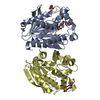 3aikS S: Starting model for refinement |
|---|---|
| Similar structure data |


-Links
 PDBj
PDBj- Assembly
| Deposited unit | 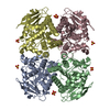
| ||||||||
|---|---|---|---|---|---|---|---|---|---|
| 1 | 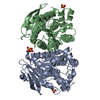
| ||||||||
| 2 | 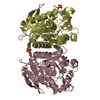
| ||||||||
| Unit cell |
| ||||||||
| Components on special symmetry positions |
|
-Components
| #1: Protein | Mass: 34540.410 Da / Num. of mol.: 4 Source method: isolated from a genetically manipulated source Details: Hyperthermophil acidophil archea Source: (gene. exp.) Sulfolobus islandicus (strain REY15A) (archaea)Strain: REY15A / Cell: Prokaryot / Cell line: E233S / Gene: SiRe_0290 / Plasmid: pSeSD Details (production host): D-Arabinose inducible promoter, shuttle vector Cell (production host): Prokaryot / Cell line (production host): E233S / Production host: Sulfolobus islandicus REY15A (archaea) / Strain (production host): SiRe_0290 / Variant (production host): REY15A / References: UniProt: F0NDQ1#2: Chemical | ChemComp-SO4 /   Mass: 96.063 Da / Num. of mol.: 8 / Source method: isolated from a natural source / Formula: SO4 Mass: 96.063 Da / Num. of mol.: 8 / Source method: isolated from a natural source / Formula: SO4#3: Water | ChemComp-HOH / |  Mass: 18.015 Da / Num. of mol.: 438 / Source method: isolated from a natural source / Formula: H2O Mass: 18.015 Da / Num. of mol.: 438 / Source method: isolated from a natural source / Formula: H2OHas protein modification | Y | |
|---|
-Experimental details
-Experiment
| Experiment | Method: X-RAY DIFFRACTION / Number of used crystals: 1 |
|---|
- Sample preparation
| Crystal | Density Matthews: 4.66 Å3/Da / Density % sol: 73.34 % / Description: Larger than 0.2 mm |
|---|---|
| Crystal grow | Temperature: 298 K / Method: vapor diffusion, hanging drop / pH: 6.5 Details: Hampton Reaserch Crystal Screen 2 (HR2-112) Condition #23. 1.6 M Ammonium sulfate, 0.1 M MES monohydrate pH 6.5, 10% v/v 1,4-Dioxane. Reservoir volume 600 uL. 2 uL of EstA 10 mg/mL in 50 mM ...Details: Hampton Reaserch Crystal Screen 2 (HR2-112) Condition #23. 1.6 M Ammonium sulfate, 0.1 M MES monohydrate pH 6.5, 10% v/v 1,4-Dioxane. Reservoir volume 600 uL. 2 uL of EstA 10 mg/mL in 50 mM Tris pH 8, mixed with 2 uL of reservoir volume for hanging drop at room temperature. PH range: 6.5 / Temp details: Room temperature |
-Data collection
| Diffraction | Mean temperature: 100 K |
|---|---|
| Diffraction source | Source: SYNCHROTRON / Site: MAX II  / Beamline: I911-3 / Wavelength: 1 Å / Beamline: I911-3 / Wavelength: 1 Å |
| Detector | Type: MARMOSAIC 225 mm CCD / Detector: CCD / Date: Oct 31, 2012 Details: First mirror: Water-cooled vertically collimating cylindrical mirror (R = 7300 m). Second mirror: Toroid mirror for horizontal and vertical focusing (R = 3300 m, R = 27 mm). |
| Radiation | Monochromator: Water-cooled double-crystal monochromator, Si(111) Protocol: SINGLE WAVELENGTH / Monochromatic (M) / Laue (L): M / Scattering type: x-ray |
| Radiation wavelength | Wavelength: 1 Å / Relative weight: 1 |
| Reflection | Resolution: 2.6→29.42 Å / Num. obs: 79041 / % possible obs: 93.3 % / Redundancy: 10.4 % / Biso Wilson estimate: 39 Å2 / Rmerge(I) obs: 0.136 / Rsym value: 0.136 / Net I/σ(I): 5.6 |
| Reflection shell | Resolution: 2.6→2.74 Å / Redundancy: 10.62 % / Rmerge(I) obs: 0.543 / Mean I/σ(I) obs: 1.4 / % possible all: 99.3 |
- Processing
| Software |
| ||||||||||||||||||||||||||||||||||||||||||||||||||||||||||||||||||||||||||||||||||||||||||||||||||||||||||||||||||||||||||||||||||||||||||||||||||||||||||||||||||||||||||||||||||||||
|---|---|---|---|---|---|---|---|---|---|---|---|---|---|---|---|---|---|---|---|---|---|---|---|---|---|---|---|---|---|---|---|---|---|---|---|---|---|---|---|---|---|---|---|---|---|---|---|---|---|---|---|---|---|---|---|---|---|---|---|---|---|---|---|---|---|---|---|---|---|---|---|---|---|---|---|---|---|---|---|---|---|---|---|---|---|---|---|---|---|---|---|---|---|---|---|---|---|---|---|---|---|---|---|---|---|---|---|---|---|---|---|---|---|---|---|---|---|---|---|---|---|---|---|---|---|---|---|---|---|---|---|---|---|---|---|---|---|---|---|---|---|---|---|---|---|---|---|---|---|---|---|---|---|---|---|---|---|---|---|---|---|---|---|---|---|---|---|---|---|---|---|---|---|---|---|---|---|---|---|---|---|---|---|
| Refinement | Method to determine structure: MOLECULAR REPLACEMENT Starting model: 3AIK Resolution: 2.6→29.42 Å / Cor.coef. Fo:Fc: 0.953 / Cor.coef. Fo:Fc free: 0.928 / SU B: 5.985 / SU ML: 0.125 / Cross valid method: THROUGHOUT / ESU R: 0.219 / ESU R Free: 0.187 / Stereochemistry target values: MAXIMUM LIKELIHOOD / Details: HYDROGENS HAVE BEEN ADDED IN THE RIDING POSITIONS
| ||||||||||||||||||||||||||||||||||||||||||||||||||||||||||||||||||||||||||||||||||||||||||||||||||||||||||||||||||||||||||||||||||||||||||||||||||||||||||||||||||||||||||||||||||||||
| Solvent computation | Ion probe radii: 0.8 Å / Shrinkage radii: 0.8 Å / VDW probe radii: 1.2 Å / Solvent model: MASK | ||||||||||||||||||||||||||||||||||||||||||||||||||||||||||||||||||||||||||||||||||||||||||||||||||||||||||||||||||||||||||||||||||||||||||||||||||||||||||||||||||||||||||||||||||||||
| Displacement parameters | Biso mean: 28.085 Å2
| ||||||||||||||||||||||||||||||||||||||||||||||||||||||||||||||||||||||||||||||||||||||||||||||||||||||||||||||||||||||||||||||||||||||||||||||||||||||||||||||||||||||||||||||||||||||
| Refinement step | Cycle: LAST / Resolution: 2.6→29.42 Å
| ||||||||||||||||||||||||||||||||||||||||||||||||||||||||||||||||||||||||||||||||||||||||||||||||||||||||||||||||||||||||||||||||||||||||||||||||||||||||||||||||||||||||||||||||||||||
| Refine LS restraints |
|