 Movie
Movie Controller

[English] 日本語
 Yorodumi
- PDB-6eo1: The electron crystallography structure of the cAMP-bound potassiu... -
+ Open data

- Basic information
| Entry | Database: PDB / ID: 6eo1 | ||||||
|---|---|---|---|---|---|---|---|
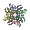
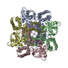
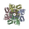
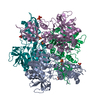
| Title | The electron crystallography structure of the cAMP-bound potassium channel MloK1 (PCO-refined) | ||||||
 Components Components | Cyclic nucleotide-gated potassium channel mll3241 | ||||||
 Keywords Keywords | MEMBRANE PROTEIN / MloK1 / MlotiK1 / potassium channel / CNBD / cytoplasmic domains / PCO refinement | ||||||
| Function / homology |  Function and homology information Function and homology informationintracellularly cyclic nucleotide-activated monoatomic cation channel activity / potassium channel activity / cAMP binding / protein-containing complex binding / identical protein binding / plasma membrane Similarity search - Function | ||||||
| Biological species |  Mesorhizobium loti MAFF303099 (bacteria) Mesorhizobium loti MAFF303099 (bacteria) | ||||||
| Method | ELECTRON CRYSTALLOGRAPHY / electron crystallography / cryo EM / Resolution: 4.5 Å | ||||||
 Authors Authors | Kowal, J. / Biyani, N. / Chami, M. / Scherer, S. / Rzepiela, A. / Baumgartner, P. / Upadhyay, V. / Nimigean, C. / Stahlberg, H. | ||||||
| Funding support |  Switzerland, 1items Switzerland, 1items
| ||||||
 Citation Citation | Journal: Structure / Year: 2018 Title: High-Resolution Cryoelectron Microscopy Structure of the Cyclic Nucleotide-Modulated Potassium Channel MloK1 in a Lipid Bilayer. Authors: Julia Kowal / Nikhil Biyani / Mohamed Chami / Sebastian Scherer / Andrzej J Rzepiela / Paul Baumgartner / Vikrant Upadhyay / Crina M Nimigean / Henning Stahlberg /  Abstract: Eukaryotic cyclic nucleotide-modulated channels perform their diverse physiological roles by opening and closing their pores to ions in response to cyclic nucleotide binding. We here present a ...Eukaryotic cyclic nucleotide-modulated channels perform their diverse physiological roles by opening and closing their pores to ions in response to cyclic nucleotide binding. We here present a structural model for the cyclic nucleotide-modulated potassium channel homolog from Mesorhizobium loti, MloK1, determined from 2D crystals in the presence of lipids. Even though crystals diffract electrons to only ∼10 Å, using cryoelectron microscopy (cryo-EM) and recently developed computational methods, we have determined a 3D map of full-length MloK1 in the presence of cyclic AMP (cAMP) at ∼4.5 Å isotropic 3D resolution. The structure provides a clear picture of the arrangement of the cyclic nucleotide-binding domains with respect to both the pore and the putative voltage sensor domains when cAMP is bound, and reveals a potential gating mechanism in the context of the lipid-embedded channel. | ||||||
| History |
|
- Structure visualization
| Movie |
Movie viewer |
|---|---|
| Structure viewer | Molecule: MolmilJmol/JSmol |
- Downloads & links
-Download
| PDBx/mmCIF format | 6eo1.cif.gz | 360.8 KB | Display | PDBx/mmCIF format |
|---|---|---|---|---|
| PDB format | pdb6eo1.ent.gz | 273.1 KB | Display | PDB format |
| PDBx/mmJSON format | 6eo1.json.gz | Tree view | PDBx/mmJSON format | |
| Others |  Other downloads Other downloads |
-Validation report
| Arichive directory | https://data.pdbj.org/pub/pdb/validation_reports/eo/6eo1ftp://data.pdbj.org/pub/pdb/validation_reports/eo/6eo1 | HTTPS FTP |
|---|
-Related structure data
| Related structure data |  3907MC M: map data used to model this data C: citing same article ( |
|---|---|
| Similar structure data |


-Links
 PDBj
PDBj


- Assembly
| Deposited unit | 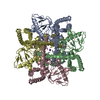
| ||||||||
|---|---|---|---|---|---|---|---|---|---|
| 1 |
| ||||||||
| Unit cell |
|
-Components
| #1: Protein | Mass: 37766.297 Da / Num. of mol.: 4 Source method: isolated from a genetically manipulated source Source: (gene. exp.) Mesorhizobium loti MAFF303099 (bacteria)Gene: mll3241 / Organ: Membrane / Plasmid: pASK90 / Cell (production host): E. coli / Production host: #2: Chemical |   Mass: 39.098 Da / Num. of mol.: 2 / Source method: obtained synthetically / Formula: K Mass: 39.098 Da / Num. of mol.: 2 / Source method: obtained synthetically / Formula: KHas protein modification | N | |
|---|
-Experimental details
-Experiment
| Experiment | Method: ELECTRON CRYSTALLOGRAPHY |
|---|---|
| EM experiment | Aggregation state: 2D ARRAY / 3D reconstruction method: electron crystallography |
| Crystal symmetry | ∠γ: 90 ° / C sampling length: 135 Å / A: 135 Å / B: 135 Å / C: 200 Å / Space group name H-M: P4212 |
- Sample preparation
| Component | Name: MloK1 tetramer / Type: COMPLEX Details: Cyclic nucleotide-modulated potassium channel in the presence of cAMP ligand, reconstituted into 2D lipid membrane crystals. Entity ID: #1 / Source: RECOMBINANT |
|---|---|
| Molecular weight | Value: 0.148 MDa / Experimental value: NO |
| Source (natural) | Organism: Mesorhizobium loti (bacteria) / Organ: Membrane |
| Source (recombinant) | Organism: |
| EM crystal formation | Instrument: dialysis buttons / Atmosphere: dialysis buffer Details: DM solubilized MloK1 sample was mixed with E. coli polar lipid extract (Avanti Polar Lipids) at a lipid to protein ratio of 0.8 and dialyzed against detergent free buffer. 2D crystals of the ...Details: DM solubilized MloK1 sample was mixed with E. coli polar lipid extract (Avanti Polar Lipids) at a lipid to protein ratio of 0.8 and dialyzed against detergent free buffer. 2D crystals of the lipid embedded protein were obtained within 5 days. Lipid mixture: E.coli polar lipids / Lipid protein ratio: 0.8 / Temperature: 293 K / Time: 5 DAY |
| Buffer solution | pH: 7.6 Details: 20 mM KCl, 20 mM Tris-HCl pH 7.6, 1 mM BaCl2, 1 mM EDTA, 0.2 mM cAMP |
| Specimen | Conc.: 0.7 mg/ml / Embedding applied: NO / Shadowing applied: NO / Staining applied: NO / Vitrification applied: YES |
| Specimen support | Grid material: COPPER / Grid mesh size: 400 divisions/in. / Grid type: Quantifoil R3.5/1 |
| Vitrification | Instrument: FEI VITROBOT MARK IV / Cryogen name: ETHANE / Humidity: 90 % / Chamber temperature: 293 K / Details: 3.5 second-blotting |
-Data collection
| Experimental equipment |  Model: Titan Krios / Image courtesy: FEI Company |
|---|---|
| Microscopy | Model: FEI TITAN KRIOS / Details: pixel size 1.3 A/pix |
| Electron gun | Electron source:  FIELD EMISSION GUN / Accelerating voltage: 300 kV / Illumination mode: FLOOD BEAM FIELD EMISSION GUN / Accelerating voltage: 300 kV / Illumination mode: FLOOD BEAM |
| Electron lens | Mode: BRIGHT FIELD / Nominal magnification: 50000 X / Nominal defocus max: 4300 nm / Nominal defocus min: 750 nm / Cs: 2.7 mm / C2 aperture diameter: 100 µm / Alignment procedure: COMA FREE |
| Specimen holder | Cryogen: NITROGEN / Specimen holder model: FEI TITAN KRIOS AUTOGRID HOLDER |
| Image recording | Average exposure time: 16 sec. / Electron dose: 45 e/Å2 / Detector mode: COUNTING / Film or detector model: GATAN K2 SUMMIT (4k x 4k) / Num. of grids imaged: 30 / Num. of real images: 346 Details: Each image was dose-fractionated in 40 frames (16 sec in total, 0.4-sec frames). The dose rate was set to ~5 counts/sec/physical-pixel (~2.8 e-/s/A2)leading to a total dose of ~45 e-/A2. Pixel size was 1.3A/pix. |
| EM imaging optics | Energyfilter name: GIF Quantum LS / Energyfilter upper: 20 eV / Energyfilter lower: 0 eV |
| Image scans | Movie frames/image: 40 |
| EM diffraction | Camera length: 800 mm / Tilt angle list: 0, 55 |
| EM diffraction shell | Resolution: 4.5→6 Å / Fourier space coverage: 100 % / Multiplicity: 300 / Num. of structure factors: 6901357 / Phase residual: 99 ° |
| EM diffraction stats | Details: Image processing was done with FOCUS (formerly 2dx), available at FOCUS-EM.org. Arheit et al., 2013a; Arheit et al., 2013b; Arheit et al., 2013c; Gipson et al., 2008; Gipson et al., 2007; ...Details: Image processing was done with FOCUS (formerly 2dx), available at FOCUS-EM.org. Arheit et al., 2013a; Arheit et al., 2013b; Arheit et al., 2013c; Gipson et al., 2008; Gipson et al., 2007; Gipson et al., 2011; Biyani et al., 2017. Rsym, Rmerge, and phase errors are not available. Fourier space coverage: 100 % / High resolution: 4.5 Å / Num. of intensities measured: 6361 / Num. of structure factors: 6901357 / Phase error: 99 ° / Phase residual: 99 ° / Phase error rejection criteria: 99 / Rmerge: 99 / Rsym: 99 |
- Processing
| Software | Name: PHENIX / Version: 1.10.1_2155: / Classification: refinement | |||||||||||||||||||||||||
|---|---|---|---|---|---|---|---|---|---|---|---|---|---|---|---|---|---|---|---|---|---|---|---|---|---|---|
| EM software |
| |||||||||||||||||||||||||
| Image processing | Details: Gatan Quantum-LS energy filter, with K2 Summit detector | |||||||||||||||||||||||||
| Crystal symmetry | ∠γ: 90 ° / C sampling length: 135 Å / A: 135 Å / B: 135 Å / C: 200 Å / Space group name H-M: P4212 | |||||||||||||||||||||||||
| CTF correction | Type: PHASE FLIPPING AND AMPLITUDE CORRECTION | |||||||||||||||||||||||||
| 3D reconstruction | Method: CRYSTALLOGRAPHY / Resolution: 4.5 Å / Resolution method: OTHER / Symmetry type: 2D CRYSTAL | |||||||||||||||||||||||||
| Atomic model building | Protocol: FLEXIBLE FIT / Space: REAL / Target criteria: fit energy Details: The initial model was obtained using Modeller (Sali and Blundell, 1993); in particular, the missing fragments were generated for the previously published PDB 4CHV model. This starting model ...Details: The initial model was obtained using Modeller (Sali and Blundell, 1993); in particular, the missing fragments were generated for the previously published PDB 4CHV model. This starting model was refined using the Rosetta for cryo-EM package (DiMaio et al., 2015). The symmetry of the channel was restrained during optimization runs (performed following the package tutorial (Wang and DiMaio,2015)). A model with a high fit score to the cryo-EM map and a low energy, as defined by the Rosetta force field, was selected from 100 Rosetta models generated and refined further. Several rounds of manual refinement with Coot (Emsley et al.,2010) and global optimization with Phenix (real_space_refine method (Afonine et al., 2013)) were carried out. Secondary structure constraints were imposed to stabilize the fold of helices and b-sheets during the global optimization. | |||||||||||||||||||||||||
| Atomic model building | PDB-ID: 4CHV Pdb chain-ID: A / Accession code: 4CHV / Pdb chain residue range: 1-355 / Source name: PDB / Type: experimental model | |||||||||||||||||||||||||
| Refinement | Resolution: 4.5→6 Å / Cross valid method: NONE /
| |||||||||||||||||||||||||
| Refine LS restraints |
|

