positive regulation of adaptive immune memory response / positive regulation of protein catabolic process in the vacuole / CD4-positive, alpha-beta T cell costimulation / osteoclast fusion / positive regulation of B cell receptor signaling pathway / myoblast fusion involved in skeletal muscle regeneration / positive regulation of T cell activation via T cell receptor contact with antigen bound to MHC molecule on antigen presenting cell / positive regulation of inflammatory response to antigenic stimulus / regulation of macrophage migration / macrophage fusion ...positive regulation of adaptive immune memory response / positive regulation of protein catabolic process in the vacuole / CD4-positive, alpha-beta T cell costimulation / osteoclast fusion / positive regulation of B cell receptor signaling pathway / myoblast fusion involved in skeletal muscle regeneration / positive regulation of T cell activation via T cell receptor contact with antigen bound to MHC molecule on antigen presenting cell / positive regulation of inflammatory response to antigenic stimulus / regulation of macrophage migration / macrophage fusion / immunological synapse formation / tetraspanin-enriched microdomain / positive regulation of T-helper 2 cell cytokine production / transferrin receptor binding / humoral immune response mediated by circulating immunoglobulin / positive regulation of protein exit from endoplasmic reticulum / MHC class II protein binding / protein localization to lysosome / positive regulation of CD4-positive, alpha-beta T cell proliferation / positive regulation of T cell receptor signaling pathway / cholesterol binding / immunological synapse / cellular response to low-density lipoprotein particle stimulus / positive regulation of B cell proliferation / positive regulation of receptor clustering / Regulation of Complement cascade / basal plasma membrane / protein localization to plasma membrane / regulation of protein stability / receptor internalization / integrin binding / Immunoregulatory interactions between a Lymphoid and a non-Lymphoid cell / MHC class II protein complex binding / virus receptor activity / vesicle / basolateral plasma membrane / positive regulation of MAPK cascade / focal adhesion / positive regulation of transcription by RNA polymerase II / extracellular exosome / membrane / plasma membrane / cytosol Similarity search - Function
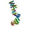
CD81antigen / 26 kDa cell surface protein TAPA-1 / Target of the antiproliferative antibody 1 / Tetraspanin-28 / Tspan-28
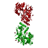
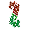
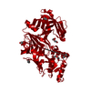
Mass: 10922.150 Da / Num. of mol.: 2 Source method: isolated from a genetically manipulated source Source: (gene. exp.) Homo sapiens (human) / Gene: CD81, TAPA1, TSPAN28 / Production host: Escherichia coli (E. coli) / References: UniProt: P60033
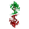
#2: Antibody
SINGLECHAINFVFRAGMENT
Mass: 26399.332 Da / Num. of mol.: 2 Source method: isolated from a genetically manipulated source Source: (gene. exp.) Mus musculus (house mouse) / Production host: Escherichia coli (E. coli)
 Movie
Movie Controller

 Yorodumi
 Open data
 Basic information
 Components
 Keywords
 Function and homology information
 Homo sapiens (human)
 X-RAY DIFFRACTION /
  Authors
 Citation
 Structure visualization
 Downloads & links
 Other downloads


 PDBj
PDBj

 Assembly

 Mass: 18.015 Da / Num. of mol.: 57 / Source method: isolated from a natural source / Formula: H2O
 Sample preparation
 / Beamline: BL9-2 / Wavelength: 0.97946 Å
 Processing