 Movie
Movie Controller

[English] 日本語
 Yorodumi
- PDB-6ehe: OmpTdeltaL8 (loop L8 deletion mutant of OmpT), an outer membrane ... -
+ Open data

- Basic information
| Entry | Database: PDB / ID: 6ehe | ||||||
|---|---|---|---|---|---|---|---|
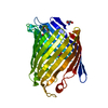
| Title | OmpTdeltaL8 (loop L8 deletion mutant of OmpT), an outer membrane protein of Vibrio cholerae | ||||||
 Components Components | OmpT protein | ||||||
 Keywords Keywords | MEMBRANE PROTEIN / Outer membrane protein / Porin / OmpF or OmpC ortholog / ion-transport / membrane beta barrel / ion-channel / diffusion porin / diffusion channel / non-specific porin. | ||||||
| Function / homology |  Function and homology information Function and homology information | ||||||
| Biological species |  Vibrio cholerae serotype O1 (bacteria) Vibrio cholerae serotype O1 (bacteria) | ||||||
| Method |  X-RAY DIFFRACTION / SYNCHROTRON / MOLECULAR REPLACEMENT / Resolution: 2.312 Å X-RAY DIFFRACTION / SYNCHROTRON / MOLECULAR REPLACEMENT / Resolution: 2.312 Å | ||||||
 Authors Authors | van den berg, B. / Pathania, M. | ||||||
| Funding support |  United Kingdom, 1items United Kingdom, 1items
| ||||||
 Citation Citation | Journal: Structure / Year: 2018 Title: Unusual Constriction Zones in the Major Porins OmpU and OmpT from Vibrio cholerae. Authors: Pathania, M. / Acosta-Gutierrez, S. / Bhamidimarri, S.P. / Basle, A. / Winterhalter, M. / Ceccarelli, M. / van den Berg, B. | ||||||
| History |
|
- Structure visualization
| Structure viewer | Molecule: MolmilJmol/JSmol |
|---|
- Downloads & links
-Download
| PDBx/mmCIF format | 6ehe.cif.gz | 77.5 KB | Display | PDBx/mmCIF format |
|---|---|---|---|---|
| PDB format | pdb6ehe.ent.gz | 56.5 KB | Display | PDB format |
| PDBx/mmJSON format | 6ehe.json.gz | Tree view | PDBx/mmJSON format | |
| Others |  Other downloads Other downloads |
-Validation report
| Arichive directory | https://data.pdbj.org/pub/pdb/validation_reports/eh/6eheftp://data.pdbj.org/pub/pdb/validation_reports/eh/6ehe | HTTPS FTP |
|---|
-Related structure data
| Related structure data |  5oykC  6ehbC  6ehcC  6ehdSC  6ehfC S: Starting model for refinement C: citing same article ( |
|---|---|
| Similar structure data |


-Links
 PDBj
PDBj- Assembly
| Deposited unit | 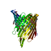
| ||||||||
|---|---|---|---|---|---|---|---|---|---|
| 1 |
| ||||||||
| Unit cell |
| ||||||||
| Components on special symmetry positions |
|
-Components
| #1: Protein | Mass: 34739.754 Da / Num. of mol.: 1 Source method: isolated from a genetically manipulated source Details: OmpTdeltaL8 is 18 residue deletion from the L8 loop of OmpT (Thr294 to Thr309, in mature sequence). Source: (gene. exp.) Vibrio cholerae serotype O1 (strain ATCC 39541 / Classical Ogawa 395 / O395) (bacteria)Gene: ompT, VC0395_A1445 / Plasmid: pET28a / Production host: | ||||||
|---|---|---|---|---|---|---|---|
| #2: Chemical | ChemComp-C8E / (   Mass: 306.438 Da / Num. of mol.: 8 / Source method: obtained synthetically / Formula: C16H34O5 / Comment: C8E, detergent*YM Mass: 306.438 Da / Num. of mol.: 8 / Source method: obtained synthetically / Formula: C16H34O5 / Comment: C8E, detergent*YM#3: Chemical | ChemComp-MG / |   Mass: 24.305 Da / Num. of mol.: 1 / Source method: obtained synthetically / Formula: Mg Mass: 24.305 Da / Num. of mol.: 1 / Source method: obtained synthetically / Formula: Mg#4: Chemical |   Mass: 59.044 Da / Num. of mol.: 2 / Source method: obtained synthetically / Formula: C2H3O2 Mass: 59.044 Da / Num. of mol.: 2 / Source method: obtained synthetically / Formula: C2H3O2#5: Water | ChemComp-HOH / |  Mass: 18.015 Da / Num. of mol.: 39 / Source method: isolated from a natural source / Formula: H2O Mass: 18.015 Da / Num. of mol.: 39 / Source method: isolated from a natural source / Formula: H2O |
-Experimental details
-Experiment
| Experiment | Method: X-RAY DIFFRACTION / Number of used crystals: 1 |
|---|
- Sample preparation
| Crystal | Density Matthews: 3.1 Å3/Da / Density % sol: 60 % |
|---|---|
| Crystal grow | Temperature: 298 K / Method: vapor diffusion, hanging drop / pH: 9.5 / Details: 0.05 magnesium acetate, 0.1 M glycine, 32% PEG 400 |
-Data collection
| Diffraction | Mean temperature: 298 K |
|---|---|
| Diffraction source | Source: SYNCHROTRON / Site: Diamond / Beamline: I24 / Wavelength: 0.9686 Å |
| Detector | Type: DECTRIS PILATUS 6M-F / Detector: PIXEL / Date: Feb 5, 2017 |
| Radiation | Protocol: SINGLE WAVELENGTH / Monochromatic (M) / Laue (L): M / Scattering type: x-ray |
| Radiation wavelength | Wavelength: 0.9686 Å / Relative weight: 1 |
| Reflection | Resolution: 2.31→85.84 Å / Num. obs: 18688 / % possible obs: 99 % / Redundancy: 6.9 % / CC1/2: 0.9 / Rpim(I) all: 0.034 / Net I/σ(I): 13.1 |
| Reflection shell | Highest resolution: 2.31 Å / Redundancy: 5.1 % / Mean I/σ(I) obs: 1.5 / CC1/2: 0.6 / Rpim(I) all: 0.51 / % possible all: 98 |
- Processing
| Software |
| |||||||||||||||||||||||||||||||||||||||||||||||||
|---|---|---|---|---|---|---|---|---|---|---|---|---|---|---|---|---|---|---|---|---|---|---|---|---|---|---|---|---|---|---|---|---|---|---|---|---|---|---|---|---|---|---|---|---|---|---|---|---|---|---|
| Refinement | Method to determine structure: MOLECULAR REPLACEMENT Starting model: 6EHD Resolution: 2.312→52.271 Å / SU ML: 0.28 / Cross valid method: FREE R-VALUE / σ(F): 1.36 / Phase error: 31.34
| |||||||||||||||||||||||||||||||||||||||||||||||||
| Solvent computation | Shrinkage radii: 0.9 Å / VDW probe radii: 1.11 Å | |||||||||||||||||||||||||||||||||||||||||||||||||
| Refinement step | Cycle: LAST / Resolution: 2.312→52.271 Å
| |||||||||||||||||||||||||||||||||||||||||||||||||
| Refine LS restraints |
| |||||||||||||||||||||||||||||||||||||||||||||||||
| LS refinement shell |
|
