National Institutes of Health/National Human Genome Research Institute (NIH/NHGRI)
R35GM122510
United States
National Institutes of Health/National Human Genome Research Institute (NIH/NHGRI)
1DP2AI138259-01
United States
National Science Foundation (NSF, United States)
1749662
United States
Citation
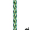
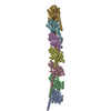
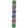
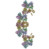
Journal: Cell / Year: 2019 Title: Structure of Microbial Nanowires Reveals Stacked Hemes that Transport Electrons over Micrometers. Authors: Fengbin Wang / Yangqi Gu / J Patrick O'Brien / Sophia M Yi / Sibel Ebru Yalcin / Vishok Srikanth / Cong Shen / Dennis Vu / Nicole L Ing / Allon I Hochbaum / Edward H Egelman / Nikhil S Malvankar / Abstract: Long-range (>10 μm) transport of electrons along networks of Geobacter sulfurreducens protein filaments, known as microbial nanowires, has been invoked to explain a wide range of globally important ...Long-range (>10 μm) transport of electrons along networks of Geobacter sulfurreducens protein filaments, known as microbial nanowires, has been invoked to explain a wide range of globally important redox phenomena. These nanowires were previously thought to be type IV pili composed of PilA protein. Here, we report a 3.7 Å resolution cryoelectron microscopy structure, which surprisingly reveals that, rather than PilA, G. sulfurreducens nanowires are assembled by micrometer-long polymerization of the hexaheme cytochrome OmcS, with hemes packed within ∼3.5-6 Å of each other. The inter-subunit interfaces show unique structural elements such as inter-subunit parallel-stacked hemes and axial coordination of heme by histidines from neighboring subunits. Wild-type OmcS filaments show 100-fold greater conductivity than other filaments from a ΔomcS strain, highlighting the importance of OmcS to conductivity in these nanowires. This structure explains the remarkable capacity of soil bacteria to transport electrons to remote electron acceptors for respiration and energy sharing.
Evidence: microscopy, helical filament was observed by negative staining and Cryo-EM
Type
Name
Symmetry operation
Number
identity operation
1_555
1
Buried area
67310 Å2
ΔGint
-1047 kcal/mol
Surface area
92910 Å2
Symmetry
Helical symmetry: (Circular symmetry: 1 / Dyad axis: no / N subunits divisor: 1 / Num. of operations: 7 / Rise per n subunits: 46.7 Å / Rotation per n subunits: -83.1 °)
-
Components
#1: Protein
C-typecytochromeOmcS / Outer membrane cytochrome S
In the structure databanks used in Yorodumi, some data are registered as the other names, "COVID-19 virus" and "2019-nCoV". Here are the details of the virus and the list of structure data.
Jan 31, 2019. EMDB accession codes are about to change! (news from PDBe EMDB page)
EMDB accession codes are about to change! (news from PDBe EMDB page)
The allocation of 4 digits for EMDB accession codes will soon come to an end. Whilst these codes will remain in use, new EMDB accession codes will include an additional digit and will expand incrementally as the available range of codes is exhausted. The current 4-digit format prefixed with “EMD-” (i.e. EMD-XXXX) will advance to a 5-digit format (i.e. EMD-XXXXX), and so on. It is currently estimated that the 4-digit codes will be depleted around Spring 2019, at which point the 5-digit format will come into force.
The EM Navigator/Yorodumi systems omit the EMD- prefix.
Related info.:Q: What is EMD? / ID/Accession-code notation in Yorodumi/EM Navigator
Yorodumi is a browser for structure data from EMDB, PDB, SASBDB, etc.
This page is also the successor to EM Navigator detail page, and also detail information page/front-end page for Omokage search.
The word "yorodu" (or yorozu) is an old Japanese word meaning "ten thousand". "mi" (miru) is to see.
Related info.:EMDB / PDB / SASBDB / Comparison of 3 databanks / Yorodumi Search / Aug 31, 2016. New EM Navigator & Yorodumi / Yorodumi Papers / Jmol/JSmol / Function and homology information / Changes in new EM Navigator and Yorodumi
 Movie
Movie Controller


 Open data
 Basic information
 Components
 Keywords
 Function and homology information
 Geobacter sulfurreducens (bacteria)
 Authors
 United States, 3items
  Citation
 Structure visualization
 Downloads & links
 Other downloads
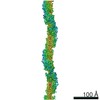


 PDBj
PDBj


 Assembly
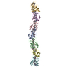

 Mass: 618.503 Da / Num. of mol.: 42 / Source method: obtained synthetically / Formula: C34H34FeN4O4
 Sample preparation
 Electron microscopy imaging
 FIELD EMISSION GUN / Accelerating voltage: 300 kV / Illumination mode: FLOOD BEAM
 Processing