 Movie
Movie Controller

[English] 日本語
 Yorodumi
- PDB-6een: Crystal structure of a designer Pentatrico Peptide RNA binding pr... -
+ Open data

- Basic information
| Entry | Database: PDB / ID: 6een | ||||||
|---|---|---|---|---|---|---|---|
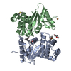
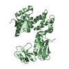
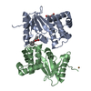
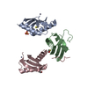
| Title | Crystal structure of a designer Pentatrico Peptide RNA binding protein, bound to a complex RNA target and featuring an infinite superhelix and microheterogeneity. | ||||||
 Components Components |
| ||||||
 Keywords Keywords | RNA BINDING PROTEIN/RNA / Complex / PPR / RNA / Helical disorder / RNA BINDING PROTEIN / RNA BINDING PROTEIN-RNA complex | ||||||
| Function / homology | RNA Function and homology information Function and homology information | ||||||
| Biological species |  | ||||||
| Method |  X-RAY DIFFRACTION / SYNCHROTRON / MOLECULAR REPLACEMENT / Resolution: 2.01 Å X-RAY DIFFRACTION / SYNCHROTRON / MOLECULAR REPLACEMENT / Resolution: 2.01 Å | ||||||
 Authors Authors | Schmidberger, J.W. / Bond, C.S. | ||||||
| Funding support |  Australia, 1items Australia, 1items
| ||||||
 Citation Citation | Journal: To Be Published Title: Crystal structure of a designer Pentatrico Peptide RNA binding protein, bound to a complex RNA target and featuring an infinite superhelix and microheterogeneity. Authors: Schmidberger, J.W. / Bond, C.S. | ||||||
| History |
|
- Structure visualization
| Structure viewer | Molecule: MolmilJmol/JSmol |
|---|
- Downloads & links
-Download
| PDBx/mmCIF format | 6een.cif.gz | 271.8 KB | Display | PDBx/mmCIF format |
|---|---|---|---|---|
| PDB format | pdb6een.ent.gz | 215.7 KB | Display | PDB format |
| PDBx/mmJSON format | 6een.json.gz | Tree view | PDBx/mmJSON format | |
| Others |  Other downloads Other downloads |
-Validation report
| Summary document | 6een_validation.pdf.gz | 480.9 KB | Display | wwPDB validaton report |
|---|---|---|---|---|
| Full document | 6een_full_validation.pdf.gz | 509.7 KB | Display | |
| Data in XML | 6een_validation.xml.gz | 45.5 KB | Display | |
| Data in CIF | 6een_validation.cif.gz | 64.4 KB | Display | |
| Arichive directory | https://data.pdbj.org/pub/pdb/validation_reports/ee/6eenftp://data.pdbj.org/pub/pdb/validation_reports/ee/6een | HTTPS FTP |
-Related structure data
| Related structure data | 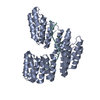 5i9fS S: Starting model for refinement |
|---|---|
| Similar structure data |
-Links
 PDBj
PDBj- Assembly
| Deposited unit | 
| ||||||||
|---|---|---|---|---|---|---|---|---|---|
| 1 |
| ||||||||
| Unit cell |
| ||||||||
| Details | this protein:RNA dimer assembly is represented as an octameric assembly due to structural disorder/microheterogeneity resulting in the structure being refined as 4 protein chains (partially occupied) + 4 RNA chains (partially occupied) |
-Components
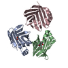
-Designer Pentatricopeptide Protein ... , 4 types, 4 molecules ABCD
| #1: Protein | Mass: 34936.477 Da / Num. of mol.: 1 Source method: isolated from a genetically manipulated source Source: (gene. exp.)  |
|---|---|
| #2: Protein | Mass: 35053.465 Da / Num. of mol.: 1 Source method: isolated from a genetically manipulated source Source: (gene. exp.) |
| #3: Protein | Mass: 34927.609 Da / Num. of mol.: 1 Source method: isolated from a genetically manipulated source Source: (gene. exp.) |
| #4: Protein | Mass: 34801.383 Da / Num. of mol.: 1 Source method: isolated from a genetically manipulated source Source: (gene. exp.) |
-RNA chain , 4 types, 4 molecules FGHI
| #5: RNA chain | Mass: 3061.895 Da / Num. of mol.: 1 / Source method: obtained synthetically / Source: (synth.) |
|---|---|
| #6: RNA chain | Mass: 2710.535 Da / Num. of mol.: 1 / Source method: obtained synthetically / Source: (synth.) |
| #7: RNA chain | Mass: 2917.895 Da / Num. of mol.: 1 / Source method: obtained synthetically / Source: (synth.) |
| #8: RNA chain | Mass: 2701.679 Da / Num. of mol.: 1 / Source method: obtained synthetically / Source: (synth.) |
-Non-polymers , 1 types, 214 molecules 
| #9: Water | ChemComp-HOH / Mass: 18.015 Da / Num. of mol.: 214 / Source method: isolated from a natural source / Formula: H2O |
|---|
-Details
| Compound details | The structure features an infinite superhelix that has inherent helical disorder. This means we ...The structure features an infinite superhelix that has inherent helical disorder. This means we don't know where it begins and ends. This leads to map averaging at each position in the repeating helix. The highly redundant design of the protein means this averaging is not an issues except at positions 5 and 35 of each repeat that change to bind different bases in the target RNA. Also, the target RNA will average out. The solution to this was to build the structure as four superposed chains for the protein and four superposed chains for the RNA. Each case adding up to an occupancy of 1. |
|---|---|
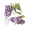
| Sequence details | Protein is designed based on a consensus sequence of many PPR proteins. Chains A to D are ...Protein is designed based on a consensus sequence of many PPR proteins. Chains A to D are repetitions of the same single chain with microheterogeneity at positions 5 and 35. The protein is observed to bind to the RNA target. The actual target sequence is GUAUCCUUAA |
-Experimental details
-Experiment
| Experiment | Method: X-RAY DIFFRACTION / Number of used crystals: 1 |
|---|
- Sample preparation
| Crystal | Density Matthews: 2.73 Å3/Da / Density % sol: 54.97 % |
|---|---|
| Crystal grow | Temperature: 293 K / Method: vapor diffusion, sitting drop / Details: 0.1 M Mg Acetate, 0.05 M MES pH 5.6, 20% MPD |
-Data collection
| Diffraction | Mean temperature: 100 K | ||||||||||||||||||||||||||||||
|---|---|---|---|---|---|---|---|---|---|---|---|---|---|---|---|---|---|---|---|---|---|---|---|---|---|---|---|---|---|---|---|
| Diffraction source | Source: SYNCHROTRON / Site: Australian Synchrotron / Beamline: MX2 / Wavelength: 0.953737 Å | ||||||||||||||||||||||||||||||
| Detector | Type: DECTRIS EIGER X 16M / Detector: PIXEL / Date: Mar 1, 2017 | ||||||||||||||||||||||||||||||
| Radiation | Protocol: SINGLE WAVELENGTH / Monochromatic (M) / Laue (L): M / Scattering type: x-ray | ||||||||||||||||||||||||||||||
| Radiation wavelength | Wavelength: 0.953737 Å / Relative weight: 1 | ||||||||||||||||||||||||||||||
| Reflection | Resolution: 2.01→44.92 Å / Num. obs: 25127 / % possible obs: 97 % / Redundancy: 2 % / CC1/2: 0.995 / Rmerge(I) obs: 0.055 / Rpim(I) all: 0.049 / Rrim(I) all: 0.074 / Net I/σ(I): 9 / Num. measured all: 50507 | ||||||||||||||||||||||||||||||
| Reflection shell | Diffraction-ID: 1
|
- Processing
| Software |
| ||||||||||||||||||||||||||||||||||||||||||||||||||||||||||||
|---|---|---|---|---|---|---|---|---|---|---|---|---|---|---|---|---|---|---|---|---|---|---|---|---|---|---|---|---|---|---|---|---|---|---|---|---|---|---|---|---|---|---|---|---|---|---|---|---|---|---|---|---|---|---|---|---|---|---|---|---|---|
| Refinement | Method to determine structure: MOLECULAR REPLACEMENT Starting model: 5i9f Resolution: 2.01→44.92 Å / Cor.coef. Fo:Fc: 0.959 / Cor.coef. Fo:Fc free: 0.928 / WRfactor Rfree: 0.2435 / WRfactor Rwork: 0.177 / FOM work R set: 0.8049 / SU B: 17.923 / SU ML: 0.477 / SU Rfree: 0.3423 / Cross valid method: THROUGHOUT / σ(F): 0 / ESU R Free: 0.342 / Stereochemistry target values: MAXIMUM LIKELIHOOD Details: HYDROGENS HAVE BEEN ADDED IN THE RIDING POSITIONS U VALUES : REFINED INDIVIDUALLY
| ||||||||||||||||||||||||||||||||||||||||||||||||||||||||||||
| Solvent computation | Ion probe radii: 0.8 Å / Shrinkage radii: 0.8 Å / VDW probe radii: 1.2 Å / Solvent model: MASK | ||||||||||||||||||||||||||||||||||||||||||||||||||||||||||||
| Displacement parameters | Biso max: 92.79 Å2 / Biso mean: 32.183 Å2 / Biso min: 11.54 Å2
| ||||||||||||||||||||||||||||||||||||||||||||||||||||||||||||
| Refinement step | Cycle: final / Resolution: 2.01→44.92 Å
| ||||||||||||||||||||||||||||||||||||||||||||||||||||||||||||
| Refine LS restraints |
| ||||||||||||||||||||||||||||||||||||||||||||||||||||||||||||
| LS refinement shell | Resolution: 2.008→2.06 Å / Rfactor Rfree error: 0 / Total num. of bins used: 20
|
