 Movie
Movie Controller

[English] 日本語
 Yorodumi
- PDB-6edu: B41 SOSIP.664 in complex with soluble CD4 (D1-D2), the co-recepto... -
+ Open data

- Basic information
| Entry | Database: PDB / ID: 6edu | ||||||||||||
|---|---|---|---|---|---|---|---|---|---|---|---|---|---|
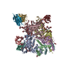
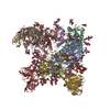
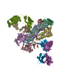
| Title | B41 SOSIP.664 in complex with soluble CD4 (D1-D2), the co-receptor mimicking antibody 21c and the broadly neutralizing antibody 8ANC195 | ||||||||||||
 Components Components |
| ||||||||||||
 Keywords Keywords | IMMUNE SYSTEM / HIV-1 Env / broadly neutralizing antibodies / cryo-EM / single particle analysis / VIRAL PROTEIN-IMMUNE SYSTEM complex | ||||||||||||
| Function / homology |  Function and homology information Function and homology informationhelper T cell enhancement of adaptive immune response / interleukin-16 binding / interleukin-16 receptor activity / cellular response to ionomycin / maintenance of protein location in cell / response to methamphetamine hydrochloride / T cell selection / MHC class II protein binding / interleukin-15-mediated signaling pathway / cellular response to granulocyte macrophage colony-stimulating factor stimulus ...helper T cell enhancement of adaptive immune response / interleukin-16 binding / interleukin-16 receptor activity / cellular response to ionomycin / maintenance of protein location in cell / response to methamphetamine hydrochloride / T cell selection / MHC class II protein binding / interleukin-15-mediated signaling pathway / cellular response to granulocyte macrophage colony-stimulating factor stimulus / positive regulation of monocyte differentiation / Alpha-defensins / Nef Mediated CD4 Down-regulation / regulation of T cell activation / response to vitamin D / Other interleukin signaling / leukocyte chemotaxis / extracellular matrix structural constituent / T cell receptor complex / enzyme-linked receptor protein signaling pathway / Translocation of ZAP-70 to Immunological synapse / Phosphorylation of CD3 and TCR zeta chains / macrophage differentiation / Generation of second messenger molecules / immunoglobulin binding / T cell differentiation / Co-inhibition by PD-1 / positive regulation of calcium ion transport into cytosol / Binding and entry of HIV virion / coreceptor activity / symbiont-mediated perturbation of host defense response / positive regulation of plasma membrane raft polarization / positive regulation of receptor clustering / positive regulation of interleukin-2 production / positive regulation of T cell proliferation / cell surface receptor protein tyrosine kinase signaling pathway / protein tyrosine kinase binding / host cell endosome membrane / T cell activation / clathrin-coated endocytic vesicle membrane / Vpu mediated degradation of CD4 / positive regulation of inflammatory response / MHC class II protein complex binding / response to estradiol / transmembrane signaling receptor activity / T cell receptor signaling pathway / Downstream TCR signaling / Cargo recognition for clathrin-mediated endocytosis / Clathrin-mediated endocytosis / virus receptor activity / signaling receptor activity / clathrin-dependent endocytosis of virus by host cell / response to ethanol / adaptive immune response / early endosome / cell surface receptor signaling pathway / cell adhesion / immune response / viral protein processing / membrane raft / endoplasmic reticulum lumen / fusion of virus membrane with host plasma membrane / external side of plasma membrane / signaling receptor binding / fusion of virus membrane with host endosome membrane / viral envelope / symbiont entry into host cell / lipid binding / protein kinase binding / endoplasmic reticulum membrane / virion attachment to host cell / host cell plasma membrane / virion membrane / structural molecule activity / enzyme binding / signal transduction / protein homodimerization activity / : / zinc ion binding / membrane / identical protein binding / plasma membrane Similarity search - Function | ||||||||||||
| Biological species |   Human immunodeficiency virus 1 Human immunodeficiency virus 1 Homo sapiens (human) Homo sapiens (human) | ||||||||||||
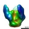
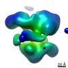
| Method | ELECTRON MICROSCOPY / single particle reconstruction / cryo EM / Resolution: 4.06 Å | ||||||||||||
 Authors Authors | Barnes, C.O. / Bjorkman, P.J. | ||||||||||||
| Funding support |  United States, 2items United States, 2items
| ||||||||||||
 Citation Citation | Journal: Cell Host Microbe / Year: 2018 Title: Partially Open HIV-1 Envelope Structures Exhibit Conformational Changes Relevant for Coreceptor Binding and Fusion. Authors: Haoqing Wang / Christopher O Barnes / Zhi Yang / Michel C Nussenzweig / Pamela J Bjorkman / Abstract: HIV-1 Env, a trimer of gp120-gp41 heterodimers, mediates membrane fusion after binding host receptor CD4. Receptor binding displaces V1V2 loops from Env's apex, allowing coreceptor binding and ...HIV-1 Env, a trimer of gp120-gp41 heterodimers, mediates membrane fusion after binding host receptor CD4. Receptor binding displaces V1V2 loops from Env's apex, allowing coreceptor binding and opening Env to enable gp41-mediated fusion. We present 3.54 Å and 4.06 Å cryoelectron microscopy structures of partially open soluble native-like Env trimers (SOSIPs) bound to CD4. One structure, a complex with a coreceptor-mimicking antibody that binds both CD4 and gp120, stabilizes the displaced V1V2 and reveals its structure. Comparing partially and fully open Envs with closed Envs shows that gp41 rearrangements are independent of the CD4-induced rearrangements that result in V1V2 displacement and formation of a 4-stranded bridging sheet. These findings suggest ordered conformational changes before coreceptor binding: (1) gp120 opening inducing side-chain rearrangements and a compact gp41 central helix conformation, and (2) 4-stranded bridging-sheet formation and V1V2 displacement. These analyses illuminate potential receptor-induced Env changes and inform design of therapeutics disrupting viral entry. | ||||||||||||
| History |
|
- Structure visualization
| Movie |
Movie viewer |
|---|---|
| Structure viewer | Molecule: MolmilJmol/JSmol |
- Downloads & links
-Download
| PDBx/mmCIF format | 6edu.cif.gz | 617.2 KB | Display | PDBx/mmCIF format |
|---|---|---|---|---|
| PDB format | pdb6edu.ent.gz | 497.3 KB | Display | PDB format |
| PDBx/mmJSON format | 6edu.json.gz | Tree view | PDBx/mmJSON format | |
| Others |  Other downloads Other downloads |
-Validation report
| Arichive directory | https://data.pdbj.org/pub/pdb/validation_reports/ed/6eduftp://data.pdbj.org/pub/pdb/validation_reports/ed/6edu | HTTPS FTP |
|---|
-Related structure data
| Related structure data | 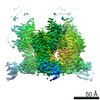 9038MC 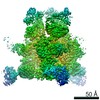 7516C 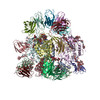 6cm3C C: citing same article ( M: map data used to model this data |
|---|---|
| Similar structure data |
-Links
 PDBj
PDBj


- Assembly
| Deposited unit | 
|
|---|---|
| 1 |
|
-Components
-Envelope glycoprotein ... , 2 types, 6 molecules ABCDEF
| #1: Protein | Mass: 17357.824 Da / Num. of mol.: 3 Source method: isolated from a genetically manipulated source Source: (gene. exp.) Human immunodeficiency virus 1 / Gene: env / Cell line (production host): CHO / Production host:   Cricetulus griseus (Chinese hamster) / Variant (production host): K1 / References: UniProt: B3UEZ6 Cricetulus griseus (Chinese hamster) / Variant (production host): K1 / References: UniProt: B3UEZ6#2: Protein | Mass: 57702.469 Da / Num. of mol.: 3 Source method: isolated from a genetically manipulated source Source: (gene. exp.) Human immunodeficiency virus 1 / Gene: env / Cell line (production host): CHO / Production host: Cricetulus griseus (Chinese hamster) / Variant (production host): K1 / References: UniProt: B3UES2 |
|---|
-Protein , 1 types, 3 molecules GHI
| #3: Protein | Mass: 20503.260 Da / Num. of mol.: 3 Source method: isolated from a genetically manipulated source Source: (gene. exp.) Homo sapiens (human) / Gene: CD4 / Cell line (production host): HEK293 / Production host: Homo sapiens (human) / References: UniProt: P01730 |
|---|
-Antibody , 4 types, 12 molecules JLNKMOPRTQSU
| #4: Antibody | Mass: 24619.590 Da / Num. of mol.: 3 Source method: isolated from a genetically manipulated source Source: (gene. exp.) Homo sapiens (human) / Cell line (production host): HEK293 / Production host: Homo sapiens (human)#5: Antibody | Mass: 22815.264 Da / Num. of mol.: 3 Source method: isolated from a genetically manipulated source Source: (gene. exp.) Homo sapiens (human) / Cell line (production host): HEK293 / Production host: Homo sapiens (human)#6: Antibody | Mass: 26125.270 Da / Num. of mol.: 3 Source method: isolated from a genetically manipulated source Source: (gene. exp.) Homo sapiens (human) / Cell line (production host): HEK293 / Production host: Homo sapiens (human)#7: Antibody | Mass: 23401.984 Da / Num. of mol.: 3 Source method: isolated from a genetically manipulated source Source: (gene. exp.) Homo sapiens (human) / Cell line (production host): HEK293 / Production host: Homo sapiens (human) |
|---|
-Sugars , 6 types, 54 molecules 
| #8: Polysaccharide | 2-acetamido-2-deoxy-beta-D-glucopyranose-(1-4)-2-acetamido-2-deoxy-beta-D-glucopyranose Source method: isolated from a genetically manipulated source #9: Polysaccharide | Source method: isolated from a genetically manipulated source #10: Polysaccharide | Source method: isolated from a genetically manipulated source #11: Polysaccharide | Source method: isolated from a genetically manipulated source #12: Polysaccharide | Source method: isolated from a genetically manipulated source #13: Sugar | ChemComp-NAG /  Type: D-saccharide, beta linking / Mass: 221.208 Da / Num. of mol.: 27 Type: D-saccharide, beta linking / Mass: 221.208 Da / Num. of mol.: 27Source method: isolated from a genetically manipulated source Formula: C8H15NO6 |
|---|
-Details
| Has protein modification | Y |
|---|
-Experimental details
-Experiment
| Experiment | Method: ELECTRON MICROSCOPY |
|---|---|
| EM experiment | Aggregation state: PARTICLE / 3D reconstruction method: single particle reconstruction |
- Sample preparation
| Component | Name: B41 SOSIP-sCD4-21c-8ANC195 complex / Type: COMPLEX / Entity ID: #1-#7 / Source: RECOMBINANT | |||||||||||||||
|---|---|---|---|---|---|---|---|---|---|---|---|---|---|---|---|---|
| Molecular weight | Value: 0.7 MDa / Experimental value: NO | |||||||||||||||
| Source (natural) | Organism: Homo sapiens (human) | |||||||||||||||
| Source (recombinant) | Organism: Homo sapiens (human) | |||||||||||||||
| Buffer solution | pH: 8 | |||||||||||||||
| Buffer component |
| |||||||||||||||
| Specimen | Conc.: 0.35 mg/ml / Embedding applied: NO / Shadowing applied: NO / Staining applied: NO / Vitrification applied: YES | |||||||||||||||
| Specimen support | Details: 0.26 mA / Grid material: COPPER / Grid mesh size: 300 divisions/in. / Grid type: Quantifoil R1.2/1.3 | |||||||||||||||
| Vitrification | Instrument: FEI VITROBOT MARK IV / Cryogen name: ETHANE / Humidity: 95 % / Chamber temperature: 293 K |
- Electron microscopy imaging
| Experimental equipment |  Model: Titan Krios / Image courtesy: FEI Company |
|---|---|
| Microscopy | Model: FEI TITAN KRIOS |
| Electron gun | Electron source:  FIELD EMISSION GUN / Accelerating voltage: 300 kV / Illumination mode: FLOOD BEAM FIELD EMISSION GUN / Accelerating voltage: 300 kV / Illumination mode: FLOOD BEAM |
| Electron lens | Mode: BRIGHT FIELD / Nominal defocus max: 3000 nm / Nominal defocus min: 1700 nm / Cs: 2.7 mm / Alignment procedure: COMA FREE |
| Specimen holder | Cryogen: NITROGEN / Specimen holder model: FEI TITAN KRIOS AUTOGRID HOLDER |
| Image recording | Average exposure time: 10 sec. / Electron dose: 60 e/Å2 / Detector mode: SUPER-RESOLUTION / Film or detector model: GATAN K2 SUMMIT (4k x 4k) / Num. of grids imaged: 1 / Num. of real images: 2531 |
| Image scans | Movie frames/image: 50 |
- Processing
| Software | Name: PHENIX / Version: 1.14_3219: / Classification: refinement | |||||||||||||||||||||||||||||||||||
|---|---|---|---|---|---|---|---|---|---|---|---|---|---|---|---|---|---|---|---|---|---|---|---|---|---|---|---|---|---|---|---|---|---|---|---|---|
| EM software |
| |||||||||||||||||||||||||||||||||||
| CTF correction | Type: PHASE FLIPPING AND AMPLITUDE CORRECTION | |||||||||||||||||||||||||||||||||||
| Symmetry | Point symmetry: C3 (3 fold cyclic) | |||||||||||||||||||||||||||||||||||
| 3D reconstruction | Resolution: 4.06 Å / Resolution method: FSC 0.143 CUT-OFF / Num. of particles: 305469 / Num. of class averages: 4 / Symmetry type: POINT | |||||||||||||||||||||||||||||||||||
| Atomic model building | Protocol: RIGID BODY FIT / Space: REAL / Target criteria: Correlation coefficient | |||||||||||||||||||||||||||||||||||
| Refinement | Highest resolution: 4.06 Å | |||||||||||||||||||||||||||||||||||
| Refine LS restraints |
|
