Movie
Movie Controller

[English] 日本語
 Yorodumi
- PDB-6ebk: The voltage-activated Kv1.2-2.1 paddle chimera channel in lipid n... -
+ Open data

- Basic information
| Entry | Database: PDB / ID: 6ebk | ||||||
|---|---|---|---|---|---|---|---|
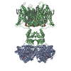
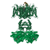
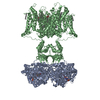
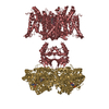
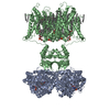
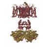
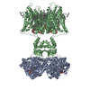
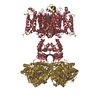
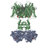
| Title | The voltage-activated Kv1.2-2.1 paddle chimera channel in lipid nanodiscs | ||||||
 Components Components |
| ||||||
 Keywords Keywords | MEMBRANE PROTEIN / transport protein / potassium channel / lipid nanodisc | ||||||
| Function / homology |  Function and homology information Function and homology informationoptic nerve structural organization / pinceau fiber / Voltage gated Potassium channels / methylglyoxal reductase (NADPH) (acetol producing) activity / voltage-gated monoatomic ion channel activity involved in regulation of postsynaptic membrane potential / potassium channel complex / regulation of circadian sleep/wake cycle, non-REM sleep / paranodal junction / potassium ion export across plasma membrane / regulation of protein localization to cell surface ...optic nerve structural organization / pinceau fiber / Voltage gated Potassium channels / methylglyoxal reductase (NADPH) (acetol producing) activity / voltage-gated monoatomic ion channel activity involved in regulation of postsynaptic membrane potential / potassium channel complex / regulation of circadian sleep/wake cycle, non-REM sleep / paranodal junction / potassium ion export across plasma membrane / regulation of protein localization to cell surface / corpus callosum development / alcohol dehydrogenase (NADP+) activity / voltage-gated monoatomic ion channel activity involved in regulation of presynaptic membrane potential / delayed rectifier potassium channel activity / axon initial segment / Oxidoreductases; Acting on the CH-OH group of donors; With NAD+ or NADP+ as acceptor / juxtaparanode region of axon / myoblast differentiation / regulation of potassium ion transmembrane transport / optic nerve development / outward rectifier potassium channel activity / Neutrophil degranulation / neuromuscular process / neuronal cell body membrane / regulation of dopamine secretion / lamellipodium membrane / action potential / potassium channel regulator activity / voltage-gated potassium channel activity / kinesin binding / hematopoietic progenitor cell differentiation / neuronal action potential / voltage-gated potassium channel complex / potassium ion transmembrane transport / sensory perception of pain / axon terminus / calyx of Held / protein localization to plasma membrane / potassium ion transport / cerebral cortex development / protein homooligomerization / postsynaptic density membrane / cytoplasmic side of plasma membrane / lamellipodium / presynaptic membrane / cytoskeleton / transmembrane transporter binding / perikaryon / postsynaptic membrane / endosome / neuron projection / postsynaptic density / protein heterodimerization activity / axon / neuronal cell body / dendrite / endoplasmic reticulum membrane / protein-containing complex binding / glutamatergic synapse / membrane / plasma membrane / cytosol Similarity search - Function | ||||||
| Biological species |  | ||||||
| Method | ELECTRON MICROSCOPY / single particle reconstruction / cryo EM / Resolution: 3.3 Å | ||||||
 Authors Authors | Matthies, D. / Bae, C. / Fox, T. / Bartesaghi, A. / Subramaniam, S. / Swartz, K.J. | ||||||
 Citation Citation | Journal: Elife / Year: 2018 Title: Single-particle cryo-EM structure of a voltage-activated potassium channel in lipid nanodiscs. Authors: Doreen Matthies / Chanhyung Bae / Gilman Es Toombes / Tara Fox / Alberto Bartesaghi / Sriram Subramaniam / Kenton Jon Swartz /  Abstract: Voltage-activated potassium (Kv) channels open to conduct K ions in response to membrane depolarization, and subsequently enter non-conducting states through distinct mechanisms of inactivation. X- ...Voltage-activated potassium (Kv) channels open to conduct K ions in response to membrane depolarization, and subsequently enter non-conducting states through distinct mechanisms of inactivation. X-ray structures of detergent-solubilized Kv channels appear to have captured an open state even though a non-conducting C-type inactivated state would predominate in membranes in the absence of a transmembrane voltage. However, structures for a voltage-activated ion channel in a lipid bilayer environment have not yet been reported. Here we report the structure of the Kv1.2-2.1 paddle chimera channel reconstituted into lipid nanodiscs using single-particle cryo-electron microscopy. At a resolution of ~3 Å for the cytosolic domain and ~4 Å for the transmembrane domain, the structure determined in nanodiscs is similar to the previously determined X-ray structure. Our findings show that large differences in structure between detergent and lipid bilayer environments are unlikely, and enable us to propose possible structural mechanisms for C-type inactivation. | ||||||
| History |
|
- Structure visualization
| Movie |
Movie viewer |
|---|---|
| Structure viewer | Molecule: MolmilJmol/JSmol |
- Downloads & links
-Download
| PDBx/mmCIF format | 6ebk.cif.gz | 511.4 KB | Display | PDBx/mmCIF format |
|---|---|---|---|---|
| PDB format | pdb6ebk.ent.gz | 406.2 KB | Display | PDB format |
| PDBx/mmJSON format | 6ebk.json.gz | Tree view | PDBx/mmJSON format | |
| Others |  Other downloads Other downloads |
-Validation report
| Arichive directory | https://data.pdbj.org/pub/pdb/validation_reports/eb/6ebkftp://data.pdbj.org/pub/pdb/validation_reports/eb/6ebk | HTTPS FTP |
|---|
-Related structure data
| Related structure data | 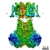 9024MC 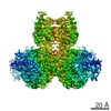 9025C  9026C 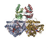 6eblC  6ebmC C: citing same article ( M: map data used to model this data |
|---|---|
| Similar structure data |

-Links
 PDBj
PDBj


- Assembly
| Deposited unit | 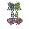
|
|---|---|
| 1 |
|
-Components
| #1: Protein | Mass: 37339.059 Da / Num. of mol.: 4 / Mutation: cytosolic domain (UNP residues 37-367) Source method: isolated from a genetically manipulated source Source: (gene. exp.)  Komagataella pastoris (fungus) / References: UniProt: P62483 Komagataella pastoris (fungus) / References: UniProt: P62483#2: Protein | Mass: 58905.828 Da / Num. of mol.: 4 Source method: isolated from a genetically manipulated source Source: (gene. exp.) Komagataella pastoris (fungus) / References: UniProt: P63142, UniProt: Q63099#3: Chemical | ChemComp-NAP /   Mass: 743.405 Da / Num. of mol.: 4 / Source method: obtained synthetically / Formula: C21H28N7O17P3 Mass: 743.405 Da / Num. of mol.: 4 / Source method: obtained synthetically / Formula: C21H28N7O17P3#4: Water | ChemComp-HOH / |  Mass: 18.015 Da / Num. of mol.: 128 / Source method: isolated from a natural source / Formula: H2O Mass: 18.015 Da / Num. of mol.: 128 / Source method: isolated from a natural source / Formula: H2O |
|---|
-Experimental details
-Experiment
| Experiment | Method: ELECTRON MICROSCOPY |
|---|---|
| EM experiment | Aggregation state: PARTICLE / 3D reconstruction method: single particle reconstruction |
- Sample preparation
| Component | Name: Voltage-activated potassium channel Kv1.2-2.1 paddle chimera in lipid nanodiscs Type: COMPLEX / Entity ID: #1-#2 / Source: RECOMBINANT | ||||||||||||||||||||
|---|---|---|---|---|---|---|---|---|---|---|---|---|---|---|---|---|---|---|---|---|---|
| Molecular weight | Value: 0.385 MDa / Experimental value: NO | ||||||||||||||||||||
| Source (natural) | Organism: | ||||||||||||||||||||
| Source (recombinant) | Organism: Komagataella pastoris (fungus) / Plasmid: pPICZ | ||||||||||||||||||||
| Buffer solution | pH: 7.5 | ||||||||||||||||||||
| Buffer component |
| ||||||||||||||||||||
| Specimen | Conc.: 0.7 mg/ml / Embedding applied: NO / Shadowing applied: NO / Staining applied: NO / Vitrification applied: YES / Details: Kv1.2-2.1 paddle chimera in lipid nanodiscs | ||||||||||||||||||||
| Specimen support | Grid material: GOLD / Grid type: Quantifoil, UltrAuFoil, R1.2/1.3 | ||||||||||||||||||||
| Vitrification | Instrument: LEICA EM GP / Cryogen name: ETHANE / Humidity: 88 % / Chamber temperature: 277.15 K Details: A 3 microliter sample was applied to a plasma-cleaned grid and blotted for 10 seconds. |
- Electron microscopy imaging
| Experimental equipment |  Model: Titan Krios / Image courtesy: FEI Company |
|---|---|
| Microscopy | Model: FEI TITAN KRIOS |
| Electron gun | Electron source:  FIELD EMISSION GUN / Accelerating voltage: 300 kV / Illumination mode: FLOOD BEAM FIELD EMISSION GUN / Accelerating voltage: 300 kV / Illumination mode: FLOOD BEAM |
| Electron lens | Mode: BRIGHT FIELD / Nominal magnification: 29000 X / Nominal defocus max: 2500 nm / Nominal defocus min: 1000 nm / Cs: 2.7 mm / C2 aperture diameter: 70 µm / Alignment procedure: COMA FREE |
| Specimen holder | Cryogen: NITROGEN / Specimen holder model: FEI TITAN KRIOS AUTOGRID HOLDER |
| Image recording | Average exposure time: 15.2 sec. / Electron dose: 40 e/Å2 / Detector mode: SUPER-RESOLUTION / Film or detector model: GATAN K2 SUMMIT (4k x 4k) / Num. of grids imaged: 1 / Num. of real images: 3085 |
| Image scans | Width: 7420 / Height: 7676 / Movie frames/image: 38 / Used frames/image: 2-20 |
- Processing
| EM software |
| ||||||||||||||||||||||||||||||||||||||||
|---|---|---|---|---|---|---|---|---|---|---|---|---|---|---|---|---|---|---|---|---|---|---|---|---|---|---|---|---|---|---|---|---|---|---|---|---|---|---|---|---|---|
| CTF correction | Type: PHASE FLIPPING ONLY | ||||||||||||||||||||||||||||||||||||||||
| Particle selection | Num. of particles selected: 281021 | ||||||||||||||||||||||||||||||||||||||||
| Symmetry | Point symmetry: C4 (4 fold cyclic) | ||||||||||||||||||||||||||||||||||||||||
| 3D reconstruction | Resolution: 3.3 Å / Resolution method: FSC 0.143 CUT-OFF / Num. of particles: 47482 / Num. of class averages: 1 / Symmetry type: POINT | ||||||||||||||||||||||||||||||||||||||||
| Atomic model building | B value: 89 / Protocol: FLEXIBLE FIT / Space: REAL | ||||||||||||||||||||||||||||||||||||||||
| Atomic model building | PDB-ID: 2R9R Accession code: 2R9R / Source name: PDB / Type: experimental model |