Movie
Movie Controller

[English] 日本語
 Yorodumi
- EMDB-9024: The voltage-activated Kv1.2-2.1 paddle chimera channel in lipid n... -
+ Open data

- Basic information
| Entry | Database: EMDB / ID: EMD-9024 | |||||||||
|---|---|---|---|---|---|---|---|---|---|---|
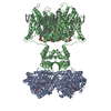
| Title | The voltage-activated Kv1.2-2.1 paddle chimera channel in lipid nanodiscs | |||||||||
 Map data Map data | Kv1.2-2.1 paddle chimera in lipid nanodiscs, entire protein | |||||||||
 Sample Sample |
| |||||||||
 Keywords Keywords | MEMBRANE PROTEIN / transport protein / potassium channel / lipid nanodisc | |||||||||
| Function / homology |  Function and homology information Function and homology informationoptic nerve structural organization / pinceau fiber / Voltage gated Potassium channels / methylglyoxal reductase (NADPH) (acetol producing) activity / voltage-gated monoatomic ion channel activity involved in regulation of postsynaptic membrane potential / potassium channel complex / regulation of circadian sleep/wake cycle, non-REM sleep / paranodal junction / potassium ion export across plasma membrane / regulation of protein localization to cell surface ...optic nerve structural organization / pinceau fiber / Voltage gated Potassium channels / methylglyoxal reductase (NADPH) (acetol producing) activity / voltage-gated monoatomic ion channel activity involved in regulation of postsynaptic membrane potential / potassium channel complex / regulation of circadian sleep/wake cycle, non-REM sleep / paranodal junction / potassium ion export across plasma membrane / regulation of protein localization to cell surface / corpus callosum development / alcohol dehydrogenase (NADP+) activity / voltage-gated monoatomic ion channel activity involved in regulation of presynaptic membrane potential / delayed rectifier potassium channel activity / axon initial segment / Oxidoreductases; Acting on the CH-OH group of donors; With NAD+ or NADP+ as acceptor / juxtaparanode region of axon / myoblast differentiation / regulation of potassium ion transmembrane transport / optic nerve development / outward rectifier potassium channel activity / Neutrophil degranulation / neuromuscular process / neuronal cell body membrane / regulation of dopamine secretion / lamellipodium membrane / action potential / potassium channel regulator activity / voltage-gated potassium channel activity / kinesin binding / hematopoietic progenitor cell differentiation / neuronal action potential / voltage-gated potassium channel complex / potassium ion transmembrane transport / sensory perception of pain / axon terminus / calyx of Held / protein localization to plasma membrane / potassium ion transport / cerebral cortex development / protein homooligomerization / postsynaptic density membrane / cytoplasmic side of plasma membrane / lamellipodium / presynaptic membrane / cytoskeleton / transmembrane transporter binding / perikaryon / postsynaptic membrane / endosome / neuron projection / postsynaptic density / protein heterodimerization activity / axon / neuronal cell body / dendrite / endoplasmic reticulum membrane / protein-containing complex binding / glutamatergic synapse / membrane / plasma membrane / cytosol Similarity search - Function | |||||||||
| Biological species |  | |||||||||
| Method | single particle reconstruction / cryo EM / Resolution: 3.3 Å | |||||||||
 Authors Authors | Matthies D / Bae C | |||||||||
 Citation Citation | Journal: Elife / Year: 2018 Title: Single-particle cryo-EM structure of a voltage-activated potassium channel in lipid nanodiscs. Authors: Doreen Matthies / Chanhyung Bae / Gilman Es Toombes / Tara Fox / Alberto Bartesaghi / Sriram Subramaniam / Kenton Jon Swartz /  Abstract: Voltage-activated potassium (Kv) channels open to conduct K ions in response to membrane depolarization, and subsequently enter non-conducting states through distinct mechanisms of inactivation. X- ...Voltage-activated potassium (Kv) channels open to conduct K ions in response to membrane depolarization, and subsequently enter non-conducting states through distinct mechanisms of inactivation. X-ray structures of detergent-solubilized Kv channels appear to have captured an open state even though a non-conducting C-type inactivated state would predominate in membranes in the absence of a transmembrane voltage. However, structures for a voltage-activated ion channel in a lipid bilayer environment have not yet been reported. Here we report the structure of the Kv1.2-2.1 paddle chimera channel reconstituted into lipid nanodiscs using single-particle cryo-electron microscopy. At a resolution of ~3 Å for the cytosolic domain and ~4 Å for the transmembrane domain, the structure determined in nanodiscs is similar to the previously determined X-ray structure. Our findings show that large differences in structure between detergent and lipid bilayer environments are unlikely, and enable us to propose possible structural mechanisms for C-type inactivation. | |||||||||
| History |
|
- Structure visualization
| Movie |
Movie viewer |
|---|---|
| Structure viewer | EM map: SurfViewMolmilJmol/JSmol |
| Supplemental images |

- Downloads & links
-EMDB archive
| Map data | emd_9024.map.gz | 132.6 MB | EMDB map data format | |
|---|---|---|---|---|
| Header (meta data) | emd-9024-v30.xmlemd-9024.xml | 19.4 KB 19.4 KB | Display Display | EMDB header |
| Images | 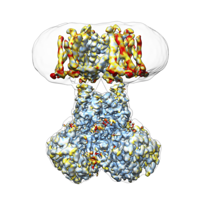 emd_9024.png emd_9024.png | 154.4 KB | ||
| Masks | emd_9024_msk_1.map | 216 MB | Mask map | |
| Filedesc metadata | emd-9024.cif.gz | 6.9 KB | ||
| Others | emd_9024_additional.map.gz | 200.8 MB | ||
| Archive directory |  http://ftp.pdbj.org/pub/emdb/structures/EMD-9024ftp://ftp.pdbj.org/pub/emdb/structures/EMD-9024 http://ftp.pdbj.org/pub/emdb/structures/EMD-9024ftp://ftp.pdbj.org/pub/emdb/structures/EMD-9024 | HTTPS FTP |
-Related structure data
| Related structure data | 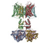 6ebkMC 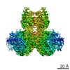 9025C  9026C 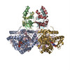 6eblC  6ebmC C: citing same article ( M: atomic model generated by this map |
|---|---|
| Similar structure data |


-Links
| EMDB pages | EMDB (EBI/PDBe) / EMDataResource |
|---|---|
| Related items in Molecule of the Month |


-Map
| File | Download / File: emd_9024.map.gz / Format: CCP4 / Size: 216 MB / Type: IMAGE STORED AS FLOATING POINT NUMBER (4 BYTES) | ||||||||||||||||||||||||||||||||||||||||||||||||||||||||||||||||||||
|---|---|---|---|---|---|---|---|---|---|---|---|---|---|---|---|---|---|---|---|---|---|---|---|---|---|---|---|---|---|---|---|---|---|---|---|---|---|---|---|---|---|---|---|---|---|---|---|---|---|---|---|---|---|---|---|---|---|---|---|---|---|---|---|---|---|---|---|---|---|
| Annotation | Kv1.2-2.1 paddle chimera in lipid nanodiscs, entire protein | ||||||||||||||||||||||||||||||||||||||||||||||||||||||||||||||||||||
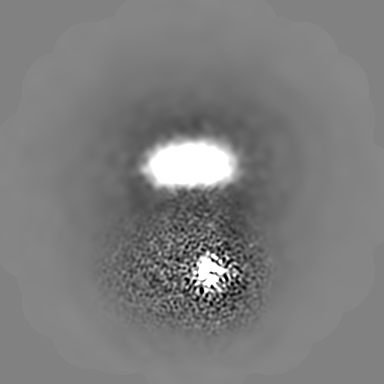
| Projections & slices | Image control
Images are generated by Spider. | ||||||||||||||||||||||||||||||||||||||||||||||||||||||||||||||||||||
| Voxel size | X=Y=Z: 0.835 Å | ||||||||||||||||||||||||||||||||||||||||||||||||||||||||||||||||||||
| Density |
| ||||||||||||||||||||||||||||||||||||||||||||||||||||||||||||||||||||
| Symmetry | Space group: 1 | ||||||||||||||||||||||||||||||||||||||||||||||||||||||||||||||||||||
| Details | EMDB XML:
CCP4 map header:
| ||||||||||||||||||||||||||||||||||||||||||||||||||||||||||||||||||||
 Z (Sec.)
 Y (Row.)
 X (Col.)


-Supplemental data
-Mask #1
| File | emd_9024_msk_1.map | ||||||||||||
|---|---|---|---|---|---|---|---|---|---|---|---|---|---|
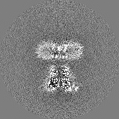
| Projections & Slices |
| ||||||||||||
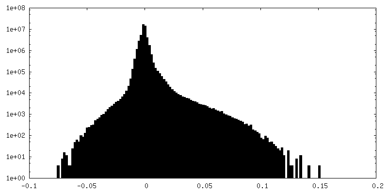
| Density Histograms |


-Additional map: Kv1.2-2.1 paddle chimera in lipid nanodiscs, additional map
| File | emd_9024_additional.map | ||||||||||||
|---|---|---|---|---|---|---|---|---|---|---|---|---|---|
| Annotation | Kv1.2-2.1 paddle chimera in lipid nanodiscs, additional map | ||||||||||||
| Projections & Slices |
| ||||||||||||
| Density Histograms |


- Sample components
-Entire : Voltage-activated potassium channel Kv1.2-2.1 paddle chimera in l...
| Entire | Name: Voltage-activated potassium channel Kv1.2-2.1 paddle chimera in lipid nanodiscs |
|---|---|
| Components |
|
-Supramolecule #1: Voltage-activated potassium channel Kv1.2-2.1 paddle chimera in l...
| Supramolecule | Name: Voltage-activated potassium channel Kv1.2-2.1 paddle chimera in lipid nanodiscs type: complex / ID: 1 / Parent: 0 / Macromolecule list: #1-#2 |
|---|---|
| Source (natural) | Organism: |
| Molecular weight | Theoretical: 385 KDa |
-Macromolecule #1: Voltage-gated potassium channel subunit beta-2
| Macromolecule | Name: Voltage-gated potassium channel subunit beta-2 / type: protein_or_peptide / ID: 1 / Number of copies: 4 / Enantiomer: LEVO |
|---|---|
| Source (natural) | Organism: |
| Molecular weight | Theoretical: 37.339059 KDa |
| Recombinant expression | Organism:  Komagataella pastoris (fungus) Komagataella pastoris (fungus) |
| Sequence | String: MVQFYRNLGK SGLRVSCLGL GTWVTFGGQI TDEMAEHLMT LAYDNGINLF DTAEVYAAGK AEVVLGNIIK KKGWRRSSLV ITTKIFWGG KAETERGLSR KHIIEGLKAS LERLQLEYVD VVFANRPDPN TPMEETVRAM THVINQGMAM YWGTSRWSSM E IMEAYSVA ...String: MVQFYRNLGK SGLRVSCLGL GTWVTFGGQI TDEMAEHLMT LAYDNGINLF DTAEVYAAGK AEVVLGNIIK KKGWRRSSLV ITTKIFWGG KAETERGLSR KHIIEGLKAS LERLQLEYVD VVFANRPDPN TPMEETVRAM THVINQGMAM YWGTSRWSSM E IMEAYSVA RQFNLIPPIC EQAEYHMFQR EKVEVQLPEL FHKIGVGAMT WSPLACGIVS GKYDSGIPPY SRASLKGYQW LK DKILSEE GRRQQAKLKE LQAIAERLGC TLPQLAIAWC LRNEGVSSVL LGASNAEQLM ENIGAIQVLP KLSSSIVHEI DSI LGNKPY SKKDYRS UniProtKB: Voltage-gated potassium channel subunit beta-2 |
-Macromolecule #2: Potassium voltage-gated channel subfamily A member 2,Potassium vo...
| Macromolecule | Name: Potassium voltage-gated channel subfamily A member 2,Potassium voltage-gated channel subfamily B member 2 chimera type: protein_or_peptide / ID: 2 / Number of copies: 4 / Enantiomer: LEVO |
|---|---|
| Source (natural) | Organism: |
| Molecular weight | Theoretical: 58.905828 KDa |
| Recombinant expression | Organism: Komagataella pastoris (fungus) |
| Sequence | String: MAHHHHHHHH ENLYFQGSMT VATGDPVDEA AAHPGHPQDT YDPEADHECC ERVVINISGL RFETQLKTLA QFPETLLGDP KKRMRYFDP LRNEYFFDRN RPSFDAILYY YQSGGRLRRP VNVPLDIFSE EIRFYELGEE AMEMFREDEG YIKEEERPLP E NEFQRQVW ...String: MAHHHHHHHH ENLYFQGSMT VATGDPVDEA AAHPGHPQDT YDPEADHECC ERVVINISGL RFETQLKTLA QFPETLLGDP KKRMRYFDP LRNEYFFDRN RPSFDAILYY YQSGGRLRRP VNVPLDIFSE EIRFYELGEE AMEMFREDEG YIKEEERPLP E NEFQRQVW LLFEYPESSG PARIIAIVSV MVILISIVSF CLETLPIFRD ENEDMHGGGV TFHTYSQSTI GYQQSTSFTD PF FIVETLC IIWFSFEFLV RFFACPSKAG FFTNIMNIID IVAIIPYYVT IFLTESNKSV LQFQNVRRVV QIFRIMRILR IFK LSRHSK GLQILGQTLK ASMRELGLLI FFLFIGVILF SSAVYFAEAD ERDSQFPSIP DAFWWAVVSM TTVGYGDMVP TTIG GKIVG SLCAIAGVLT IALPVPVIVS NFNYFYHRET EGEEQAQYLQ VTSCPKIPSS PDLKKSRSAS TISKSDYMEI QEGVN NSNE DFREENLKTA NCTLANTNYV NITKMLTDV UniProtKB: Potassium voltage-gated channel subfamily A member 2, Potassium voltage-gated channel subfamily B member 2, Potassium voltage-gated channel subfamily A member 2 |
-Macromolecule #3: NADP NICOTINAMIDE-ADENINE-DINUCLEOTIDE PHOSPHATE
| Macromolecule | Name: NADP NICOTINAMIDE-ADENINE-DINUCLEOTIDE PHOSPHATE / type: ligand / ID: 3 / Number of copies: 4 / Formula: NAP |
|---|---|
| Molecular weight | Theoretical: 743.405 Da |
| Chemical component information |  ChemComp-NAP: |
-Macromolecule #4: water
| Macromolecule | Name: water / type: ligand / ID: 4 / Number of copies: 128 / Formula: HOH |
|---|---|
| Molecular weight | Theoretical: 18.015 Da |
| Chemical component information |  ChemComp-HOH: |
-Experimental details
-Structure determination
| Method | cryo EM |
|---|---|
 Processing Processing | single particle reconstruction |
| Aggregation state | particle |
-Sample preparation
| Concentration | 0.7 mg/mL | ||||||||||||
|---|---|---|---|---|---|---|---|---|---|---|---|---|---|
| Buffer | pH: 7.5 Component:
| ||||||||||||
| Grid | Model: Quantifoil, UltrAuFoil, R1.2/1.3 / Material: GOLD / Pretreatment - Type: PLASMA CLEANING | ||||||||||||
| Vitrification | Cryogen name: ETHANE / Chamber humidity: 88 % / Chamber temperature: 277.15 K / Instrument: LEICA EM GP Details: A 3 microliter sample was applied to a plasma-cleaned grid and blotted for 10 seconds.. | ||||||||||||
| Details | Kv1.2-2.1 paddle chimera in lipid nanodiscs |
- Electron microscopy
| Microscope | FEI TITAN KRIOS |
|---|---|
| Image recording | Film or detector model: GATAN K2 SUMMIT (4k x 4k) / Detector mode: SUPER-RESOLUTION / Digitization - Dimensions - Width: 7420 pixel / Digitization - Dimensions - Height: 7676 pixel / Digitization - Frames/image: 2-20 / Number grids imaged: 1 / Number real images: 3085 / Average exposure time: 15.2 sec. / Average electron dose: 40.0 e/Å2 |
| Electron beam | Acceleration voltage: 300 kV / Electron source:  FIELD EMISSION GUN FIELD EMISSION GUN |
| Electron optics | C2 aperture diameter: 70.0 µm / Illumination mode: FLOOD BEAM / Imaging mode: BRIGHT FIELD / Cs: 2.7 mm / Nominal defocus max: 2.5 µm / Nominal defocus min: 1.0 µm / Nominal magnification: 29000 |
| Sample stage | Specimen holder model: FEI TITAN KRIOS AUTOGRID HOLDER / Cooling holder cryogen: NITROGEN |
| Experimental equipment |  Model: Titan Krios / Image courtesy: FEI Company |