 Movie
Movie Controller

[English] 日本語
 Yorodumi
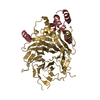
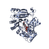
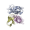
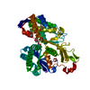
- PDB-6dxp: The crystal structure of an FMN-dependent NADH-azoreductase from ... -
+ Open data

- Basic information
| Entry | Database: PDB / ID: 6dxp | ||||||
|---|---|---|---|---|---|---|---|
| Title | The crystal structure of an FMN-dependent NADH-azoreductase from Klebsiella pneumoniae | ||||||
 Components Components | FMN-dependent NADH-azoreductase | ||||||
 Keywords Keywords | OXIDOREDUCTASE / azoreductase | ||||||
| Function / homology |  Function and homology information Function and homology informationFMN-dependent NADH-azoreductase / oxidoreductase activity, acting on NAD(P)H as acceptor / Oxidoreductases; Acting on NADH or NADPH; With a quinone or similar compound as acceptor / oxidoreductase activity, acting on NAD(P)H, quinone or similar compound as acceptor / FMN binding / electron transfer activity Similarity search - Function | ||||||
| Biological species |  Klebsiella pneumoniae (bacteria) Klebsiella pneumoniae (bacteria) | ||||||
| Method |  X-RAY DIFFRACTION / SYNCHROTRON / MOLECULAR REPLACEMENT / molecular replacement / Resolution: 2.478 Å X-RAY DIFFRACTION / SYNCHROTRON / MOLECULAR REPLACEMENT / molecular replacement / Resolution: 2.478 Å | ||||||
 Authors Authors | Arcinas, A.J. / Ghosh, A. / Chamala, S. / Bonanno, J.B. / Kelly, L. / Almo, S.C. | ||||||
 Citation Citation | Journal: To Be Published Title: The crystal structure of an FMN-dependent NADH-azoreductase from Klebsiella pneumoniae Authors: Arcinas, A.J. / Ghosh, A. / Chamala, S. / Bonanno, J.B. / Kelly, L. / Almo, S.C. | ||||||
| History |
|
- Structure visualization
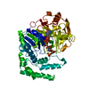
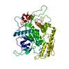
| Structure viewer | Molecule: MolmilJmol/JSmol |
|---|
- Downloads & links
-Download
| PDBx/mmCIF format | 6dxp.cif.gz | 167.7 KB | Display | PDBx/mmCIF format |
|---|---|---|---|---|
| PDB format | pdb6dxp.ent.gz | 130.1 KB | Display | PDB format |
| PDBx/mmJSON format | 6dxp.json.gz | Tree view | PDBx/mmJSON format | |
| Others |  Other downloads Other downloads |
-Validation report
| Arichive directory | https://data.pdbj.org/pub/pdb/validation_reports/dx/6dxpftp://data.pdbj.org/pub/pdb/validation_reports/dx/6dxp | HTTPS FTP |
|---|
-Related structure data
| Related structure data |  1v4bS S: Starting model for refinement |
|---|---|
| Similar structure data |


-Links
 PDBj
PDBj- Assembly
| Deposited unit | 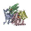
| ||||||||
|---|---|---|---|---|---|---|---|---|---|
| 1 | 
| ||||||||
| 2 | 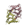
| ||||||||
| Unit cell |
|
-Components
| #1: Protein | Mass: 24257.512 Da / Num. of mol.: 4 Source method: isolated from a genetically manipulated source Source: (gene. exp.) Klebsiella pneumoniae (bacteria) / Gene: azoR, B1727_12130, C3F39_21155 / Plasmid: modified pNIC28-Bsa4 / Production host: References: UniProt: A0A1W1JPX6, UniProt: A6TCS9*PLUS, Oxidoreductases; Acting on other nitrogenous compounds as donors #2: Chemical | ChemComp-FMN /   Mass: 456.344 Da / Num. of mol.: 4 / Source method: obtained synthetically / Formula: C17H21N4O9P Mass: 456.344 Da / Num. of mol.: 4 / Source method: obtained synthetically / Formula: C17H21N4O9P#3: Water | ChemComp-HOH / |  Mass: 18.015 Da / Num. of mol.: 21 / Source method: isolated from a natural source / Formula: H2O Mass: 18.015 Da / Num. of mol.: 21 / Source method: isolated from a natural source / Formula: H2O |
|---|
-Experimental details
-Experiment
| Experiment | Method: X-RAY DIFFRACTION / Number of used crystals: 1 |
|---|
- Sample preparation
| Crystal | Density Matthews: 2.6 Å3/Da / Density % sol: 52.67 % |
|---|---|
| Crystal grow | Temperature: 291 K / Method: vapor diffusion / pH: 6 Details: 0.1M MES pH 6.0, 25% PEG 8000, 0.2M calcium acetate |
-Data collection
| Diffraction | Mean temperature: 100 K | ||||||||||||||||||||||||
|---|---|---|---|---|---|---|---|---|---|---|---|---|---|---|---|---|---|---|---|---|---|---|---|---|---|
| Diffraction source | Source: SYNCHROTRON / Site: APS  / Beamline: 31-ID / Wavelength: 0.97931 Å / Beamline: 31-ID / Wavelength: 0.97931 Å | ||||||||||||||||||||||||
| Detector | Type: MARMOSAIC 225 mm CCD / Detector: CCD / Date: Feb 7, 2018 | ||||||||||||||||||||||||
| Radiation | Monochromator: diamond / Protocol: SINGLE WAVELENGTH / Monochromatic (M) / Laue (L): M / Scattering type: x-ray | ||||||||||||||||||||||||
| Radiation wavelength | Wavelength: 0.97931 Å / Relative weight: 1 | ||||||||||||||||||||||||
| Reflection | Resolution: 2.478→29.17 Å / Num. obs: 34876 / % possible obs: 98.6 % / Redundancy: 3.7 % / Biso Wilson estimate: 51 Å2 / CC1/2: 0.996 / Rmerge(I) obs: 0.1 / Rpim(I) all: 0.06 / Rrim(I) all: 0.117 / Net I/σ(I): 5.6 | ||||||||||||||||||||||||
| Reflection shell | Diffraction-ID: 1
|
-Phasing
| Phasing | Method: molecular replacement |
|---|
- Processing
| Software |
| ||||||||||||||||||||||||||||||||||||||||||||||||||||||||||||
|---|---|---|---|---|---|---|---|---|---|---|---|---|---|---|---|---|---|---|---|---|---|---|---|---|---|---|---|---|---|---|---|---|---|---|---|---|---|---|---|---|---|---|---|---|---|---|---|---|---|---|---|---|---|---|---|---|---|---|---|---|---|
| Refinement | Method to determine structure: MOLECULAR REPLACEMENT Starting model: 1V4B Resolution: 2.478→20 Å / Cor.coef. Fo:Fc: 0.928 / Cor.coef. Fo:Fc free: 0.903 / SU B: 18.897 / SU ML: 0.394 / SU R Cruickshank DPI: 0.5246 / Cross valid method: THROUGHOUT / σ(F): 0 / ESU R: 0.525 / ESU R Free: 0.313 Details: HYDROGENS HAVE BEEN ADDED IN THE RIDING POSITIONS, U VALUES REFINED INDIVIDUALLY
| ||||||||||||||||||||||||||||||||||||||||||||||||||||||||||||
| Solvent computation | Ion probe radii: 0.8 Å / Shrinkage radii: 0.8 Å / VDW probe radii: 1.2 Å | ||||||||||||||||||||||||||||||||||||||||||||||||||||||||||||
| Displacement parameters | Biso max: 125.8 Å2 / Biso mean: 48.043 Å2 / Biso min: 14.42 Å2
| ||||||||||||||||||||||||||||||||||||||||||||||||||||||||||||
| Refinement step | Cycle: final / Resolution: 2.478→20 Å
| ||||||||||||||||||||||||||||||||||||||||||||||||||||||||||||
| Refine LS restraints |
| ||||||||||||||||||||||||||||||||||||||||||||||||||||||||||||
| LS refinement shell | Resolution: 2.478→2.541 Å
|
