 Movie
Movie Controller


+ Open data

- Basic information
| Entry | Database: PDB / ID: 6dxo | |||||||||
|---|---|---|---|---|---|---|---|---|---|---|
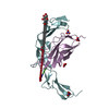
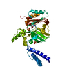
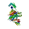
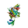
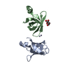
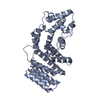
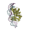
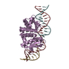
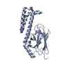
| Title | 1.8 A structure of RsbN-BldN complex. | |||||||||
 Components Components |
| |||||||||
 Keywords Keywords | TRANSCRIPTION / BldN / RsbN / Sigma / anti-sigma / ECF | |||||||||
| Function / homology |  Function and homology information Function and homology informationsigma factor activity / DNA-templated transcription initiation / DNA binding / membrane Similarity search - Function | |||||||||
| Biological species |  Streptomyces venezuelae (bacteria) Streptomyces venezuelae (bacteria) | |||||||||
| Method |  X-RAY DIFFRACTION / SYNCHROTRON / MOLECULAR REPLACEMENT / molecular replacement / Resolution: 1.8 Å X-RAY DIFFRACTION / SYNCHROTRON / MOLECULAR REPLACEMENT / molecular replacement / Resolution: 1.8 Å | |||||||||
 Authors Authors | Schumacher, M.A. | |||||||||
 Citation Citation | Journal: Nucleic Acids Res. / Year: 2018 Title: The crystal structure of the RsbN-sigma BldN complex from Streptomyces venezuelae defines a new structural class of anti-sigma factor. Authors: Schumacher, M.A. / Bush, M.J. / Bibb, M.J. / Ramos-Leon, F. / Chandra, G. / Zeng, W. / Buttner, M.J. | |||||||||
| History |
|
- Structure visualization
| Structure viewer | Molecule: MolmilJmol/JSmol |
|---|
- Downloads & links
-Download
| PDBx/mmCIF format | 6dxo.cif.gz | 98.2 KB | Display | PDBx/mmCIF format |
|---|---|---|---|---|
| PDB format | pdb6dxo.ent.gz | 73 KB | Display | PDB format |
| PDBx/mmJSON format | 6dxo.json.gz | Tree view | PDBx/mmJSON format | |
| Others |  Other downloads Other downloads |
-Validation report
| Arichive directory | https://data.pdbj.org/pub/pdb/validation_reports/dx/6dxoftp://data.pdbj.org/pub/pdb/validation_reports/dx/6dxo | HTTPS FTP |
|---|
-Related structure data
| Related structure data |  6c03 S: Starting model for refinement |
|---|---|
| Similar structure data |

-Links
 PDBj
PDBj
- Assembly
| Deposited unit | 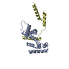
| ||||||||
|---|---|---|---|---|---|---|---|---|---|
| 1 |
| ||||||||
| Unit cell |
|
-Components
| #1: Protein | Mass: 17165.463 Da / Num. of mol.: 1 Source method: isolated from a genetically manipulated source Source: (gene. exp.) Streptomyces venezuelae (strain ATCC 10712 / CBS 650.69 / DSM 40230 / JCM 4526 / NBRC 13096 / PD 04745) (bacteria)Strain: ATCC 10712 / CBS 650.69 / DSM 40230 / JCM 4526 / NBRC 13096 / PD 04745 Gene: SVEN_3185 / Production host: Escherichia coli / References: UniProt: F2R911*PLUS |
|---|---|
| #2: Protein | Mass: 9446.593 Da / Num. of mol.: 1 Source method: isolated from a genetically manipulated source Source: (gene. exp.) Streptomyces venezuelae (strain ATCC 10712 / CBS 650.69 / DSM 40230 / JCM 4526 / NBRC 13096 / PD 04745) (bacteria)Strain: ATCC 10712 / CBS 650.69 / DSM 40230 / JCM 4526 / NBRC 13096 / PD 04745 Gene: SVEN_3186 / Production host: Escherichia coli / References: UniProt: F2R912 |
| #3: Water | ChemComp-HOH /  Mass: 18.015 Da / Num. of mol.: 135 / Source method: isolated from a natural source / Formula: H2O Mass: 18.015 Da / Num. of mol.: 135 / Source method: isolated from a natural source / Formula: H2O |
-Experimental details
-Experiment
| Experiment | Method: X-RAY DIFFRACTION / Number of used crystals: 1 |
|---|
- Sample preparation
| Crystal | Density Matthews: 1.97 Å3/Da / Density % sol: 37.58 % |
|---|---|
| Crystal grow | Temperature: 298 K / Method: vapor diffusion, hanging drop / Details: 0.2 M ammonium nitrate, 26% PEG 3500 |
-Data collection
| Diffraction | Mean temperature: 100 K |
|---|---|
| Diffraction source | Source: SYNCHROTRON / Site: ALS  / Beamline: 8.3.1 / Wavelength: 1.01 Å / Beamline: 8.3.1 / Wavelength: 1.01 Å |
| Detector | Type: ADSC QUANTUM 210r / Detector: CCD / Date: Apr 25, 2018 |
| Radiation | Protocol: SINGLE WAVELENGTH / Monochromatic (M) / Laue (L): M / Scattering type: x-ray |
| Radiation wavelength | Wavelength: 1.01 Å / Relative weight: 1 |
| Reflection | Resolution: 1.8→23.06 Å / Num. obs: 29209 / % possible obs: 92.8 % / Observed criterion σ(F): 0 / Redundancy: 2.5 % / Biso Wilson estimate: 21 Å2 / CC1/2: 0.996 / Rpim(I) all: 0.044 / Rrim(I) all: 0.075 / Rsym value: 0.06 / Net I/σ(I): 10 |
| Reflection shell | Resolution: 1.8→1.91 Å / Redundancy: 2.6 % / Mean I/σ(I) obs: 3.9 / CC1/2: 0.977 / Rpim(I) all: 0.101 / Rrim(I) all: 0.17 / Rsym value: 0.136 / % possible all: 99.3 |
-Phasing
| Phasing | Method: molecular replacement |
|---|
- Processing
| Software |
| |||||||||||||||||||||||||||||||||||||||||||||||||||||||||||||||||||||||||||||
|---|---|---|---|---|---|---|---|---|---|---|---|---|---|---|---|---|---|---|---|---|---|---|---|---|---|---|---|---|---|---|---|---|---|---|---|---|---|---|---|---|---|---|---|---|---|---|---|---|---|---|---|---|---|---|---|---|---|---|---|---|---|---|---|---|---|---|---|---|---|---|---|---|---|---|---|---|---|---|
| Refinement | Method to determine structure: MOLECULAR REPLACEMENT Starting model: 6C03 6c03 Resolution: 1.8→23.06 Å / FOM work R set: 0.82 / SU ML: 0.24 / Cross valid method: THROUGHOUT / σ(F): 0.77 / Phase error: 24.92 / Stereochemistry target values: ML
| |||||||||||||||||||||||||||||||||||||||||||||||||||||||||||||||||||||||||||||
| Solvent computation | Shrinkage radii: 0.72 Å / VDW probe radii: 1 Å / Solvent model: FLAT BULK SOLVENT MODEL / Bsol: 71.058 Å2 / ksol: 0.453 e/Å3 | |||||||||||||||||||||||||||||||||||||||||||||||||||||||||||||||||||||||||||||
| Displacement parameters | Biso max: 143.16 Å2 / Biso mean: 38.4 Å2 / Biso min: 11.45 Å2
| |||||||||||||||||||||||||||||||||||||||||||||||||||||||||||||||||||||||||||||
| Refinement step | Cycle: final / Resolution: 1.8→23.06 Å
| |||||||||||||||||||||||||||||||||||||||||||||||||||||||||||||||||||||||||||||
| Refine LS restraints |
| |||||||||||||||||||||||||||||||||||||||||||||||||||||||||||||||||||||||||||||
| LS refinement shell | Refine-ID: X-RAY DIFFRACTION / Rfactor Rfree error: 0 / Total num. of bins used: 10
| |||||||||||||||||||||||||||||||||||||||||||||||||||||||||||||||||||||||||||||
| Refinement TLS params. | Method: refined / Origin x: -5.5551 Å / Origin y: 0.569 Å / Origin z: 24.9492 Å
| |||||||||||||||||||||||||||||||||||||||||||||||||||||||||||||||||||||||||||||
| Refinement TLS group |
|
