 Movie
Movie Controller

[English] 日本語
 Yorodumi
- PDB-1li7: Crystal Structure of Cysteinyl-tRNA Synthetase with Cysteine Subs... -
+ Open data

- Basic information
| Entry | Database: PDB / ID: 1li7 | ||||||
|---|---|---|---|---|---|---|---|
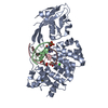
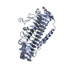
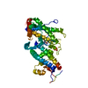
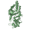
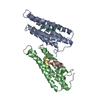
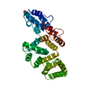
| Title | Crystal Structure of Cysteinyl-tRNA Synthetase with Cysteine Substrate Bound | ||||||
 Components Components | CYSTEINYL-TRNA SYNTHETASE | ||||||
 Keywords Keywords | LIGASE / TRNA SYNTHETASE / CYSTEINE / E.COLI | ||||||
| Function / homology |  Function and homology information Function and homology informationcysteine-tRNA ligase / cysteine-tRNA ligase activity / cysteinyl-tRNA aminoacylation / aminoacyl-tRNA ligase activity / ligase activity / zinc ion binding / ATP binding / metal ion binding / cytoplasm / cytosol Similarity search - Function | ||||||
| Biological species |  | ||||||
| Method |  X-RAY DIFFRACTION / SYNCHROTRON / FOURIER SYNTHESIS / Resolution: 2.6 Å X-RAY DIFFRACTION / SYNCHROTRON / FOURIER SYNTHESIS / Resolution: 2.6 Å | ||||||
 Authors Authors | Newberry, K.J. / Hou, Y.-M. / Perona, J.J. | ||||||
 Citation Citation | Journal: EMBO J. / Year: 2002 Title: Structural origins of amino acid selection without editing by cysteinyl-tRNA synthetase Authors: Newberry, K.J. / Hou, Y.-M. / Perona, J.J. #1: Journal: Acta Crystallogr.,Sect.D / Year: 1999Title: Crystallization and Preliminary Diffraction Analysis of Escherichia Coli Cysteinyl-tRNA Synthetase Authors: Newberry, K.J. / Kohn, J. / Hou, Y.-M. / Perona, J.J. | ||||||
| History |
|
- Structure visualization
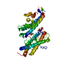
| Structure viewer | Molecule: MolmilJmol/JSmol |
|---|
- Downloads & links
-Download
| PDBx/mmCIF format | 1li7.cif.gz | 159.4 KB | Display | PDBx/mmCIF format |
|---|---|---|---|---|
| PDB format | pdb1li7.ent.gz | 122.7 KB | Display | PDB format |
| PDBx/mmJSON format | 1li7.json.gz | Tree view | PDBx/mmJSON format | |
| Others |  Other downloads Other downloads |
-Validation report
| Arichive directory | https://data.pdbj.org/pub/pdb/validation_reports/li/1li7ftp://data.pdbj.org/pub/pdb/validation_reports/li/1li7 | HTTPS FTP |
|---|
-Related structure data
| Related structure data | 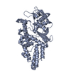 1li5SC S: Starting model for refinement C: citing same article ( |
|---|---|
| Similar structure data |

-Links
 PDBj
PDBj


- Assembly
| Deposited unit | 
| ||||||||||
|---|---|---|---|---|---|---|---|---|---|---|---|
| 1 | 
| ||||||||||
| 2 | 
| ||||||||||
| Unit cell |
| ||||||||||
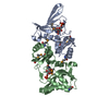
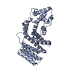
| Details | The biological unit is a monomer with one cysteine molecule bound |
-Components
| #1: Protein | Mass: 52271.059 Da / Num. of mol.: 2 Source method: isolated from a genetically manipulated source Source: (gene. exp.) #2: Chemical |   Mass: 65.409 Da / Num. of mol.: 2 / Source method: obtained synthetically / Formula: Zn Mass: 65.409 Da / Num. of mol.: 2 / Source method: obtained synthetically / Formula: Zn#3: Chemical |   Type: L-peptide linking / Mass: 121.158 Da / Num. of mol.: 2 / Source method: obtained synthetically / Formula: C3H7NO2S Type: L-peptide linking / Mass: 121.158 Da / Num. of mol.: 2 / Source method: obtained synthetically / Formula: C3H7NO2S#4: Water | ChemComp-HOH / |  Mass: 18.015 Da / Num. of mol.: 71 / Source method: isolated from a natural source / Formula: H2O Mass: 18.015 Da / Num. of mol.: 71 / Source method: isolated from a natural source / Formula: H2O |
|---|
-Experimental details
-Experiment
| Experiment | Method: X-RAY DIFFRACTION / Number of used crystals: 1 |
|---|
- Sample preparation
| Crystal | Density Matthews: 2.43 Å3/Da / Density % sol: 47 % | ||||||||||||||||||||||||||||||||||||||||||||||||||||||||||||||||||||||||||||||||||||
|---|---|---|---|---|---|---|---|---|---|---|---|---|---|---|---|---|---|---|---|---|---|---|---|---|---|---|---|---|---|---|---|---|---|---|---|---|---|---|---|---|---|---|---|---|---|---|---|---|---|---|---|---|---|---|---|---|---|---|---|---|---|---|---|---|---|---|---|---|---|---|---|---|---|---|---|---|---|---|---|---|---|---|---|---|---|
| Crystal grow | Temperature: 290 K / Method: vapor diffusion, hanging drop / pH: 6.5 Details: 16% PEG 8000, 200 MM magnesium acetate, 1MM DTT, cacodylate, 5MM ATP, 10MM cysteine. 100 MM BUFFER, pH 6.5, VAPOR DIFFUSION, HANGING DROP at 290K | ||||||||||||||||||||||||||||||||||||||||||||||||||||||||||||||||||||||||||||||||||||
| Crystal grow | *PLUS Temperature: 17 ℃ / pH: 7.4 | ||||||||||||||||||||||||||||||||||||||||||||||||||||||||||||||||||||||||||||||||||||
| Components of the solutions | *PLUS
|
-Data collection
| Diffraction | Mean temperature: 100 K |
|---|---|
| Diffraction source | Source: SYNCHROTRON / Site: NSLS  / Beamline: X12C / Wavelength: 0.98 / Wavelength: 0.98 Å / Beamline: X12C / Wavelength: 0.98 / Wavelength: 0.98 Å |
| Detector | Type: CUSTOM-MADE / Detector: CCD / Date: Sep 6, 2001 |
| Radiation | Monochromator: DOUBLE CRYSTAL MONOCHROMATOR / Protocol: SINGLE WAVELENGTH / Monochromatic (M) / Laue (L): M / Scattering type: x-ray |
| Radiation wavelength | Wavelength: 0.98 Å / Relative weight: 1 |
| Reflection | Resolution: 2.6→55 Å / Num. all: 32383 / Num. obs: 32373 / % possible obs: 100 % / Observed criterion σ(F): 0 / Observed criterion σ(I): 0 / Redundancy: 8 % / Biso Wilson estimate: 41.6 Å2 / Rmerge(I) obs: 0.091 / Net I/σ(I): 15.6 |
| Reflection shell | Resolution: 2.6→2.74 Å / Redundancy: 8.1 % / Rmerge(I) obs: 0.52 / Mean I/σ(I) obs: 4.1 / Num. unique all: 4634 / % possible all: 100 |
| Reflection | *PLUS Highest resolution: 2.6 Å / Lowest resolution: 55 Å / Rmerge(I) obs: 0.1 |
- Processing
| Software |
| ||||||||||||||||||||||||||||||||||||
|---|---|---|---|---|---|---|---|---|---|---|---|---|---|---|---|---|---|---|---|---|---|---|---|---|---|---|---|---|---|---|---|---|---|---|---|---|---|
| Refinement | Method to determine structure: FOURIER SYNTHESIS Starting model: PDB ENTRY 1LI5 Resolution: 2.6→19.95 Å / Rfactor Rfree error: 0.005 / Data cutoff high absF: 2482879.95 / Data cutoff low absF: 0 / Isotropic thermal model: RESTRAINED / Cross valid method: THROUGHOUT / σ(F): 0 / Stereochemistry target values: Engh & Huber
| ||||||||||||||||||||||||||||||||||||
| Solvent computation | Solvent model: FLAT MODEL / Bsol: 30.4565 Å2 / ksol: 0.317251 e/Å3 | ||||||||||||||||||||||||||||||||||||
| Displacement parameters | Biso mean: 40.2 Å2
| ||||||||||||||||||||||||||||||||||||
| Refine analyze |
| ||||||||||||||||||||||||||||||||||||
| Refinement step | Cycle: LAST / Resolution: 2.6→19.95 Å
| ||||||||||||||||||||||||||||||||||||
| Refine LS restraints |
| ||||||||||||||||||||||||||||||||||||
| LS refinement shell | Resolution: 2.6→2.76 Å / Rfactor Rfree error: 0.016 / Total num. of bins used: 6
| ||||||||||||||||||||||||||||||||||||
| Xplor file |
| ||||||||||||||||||||||||||||||||||||
| Refinement | *PLUS Highest resolution: 2.6 Å / Rfactor obs: 0.249 / Rfactor Rfree: 0.289 / Rfactor Rwork: 0.249 | ||||||||||||||||||||||||||||||||||||
| Solvent computation | *PLUS | ||||||||||||||||||||||||||||||||||||
| Displacement parameters | *PLUS | ||||||||||||||||||||||||||||||||||||
| Refine LS restraints | *PLUS
| ||||||||||||||||||||||||||||||||||||
| LS refinement shell | *PLUS Rfactor Rwork: 0.32 |
