 Movie
Movie Controller

[English] 日本語
 Yorodumi
- PDB-1o8b: Structure of Escherichia coli ribose-5-phosphate isomerase, RpiA,... -
+ Open data

- Basic information
| Entry | Database: PDB / ID: 1o8b | |||||||||
|---|---|---|---|---|---|---|---|---|---|---|
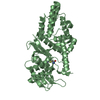
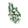
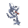
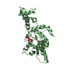
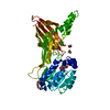
| Title | Structure of Escherichia coli ribose-5-phosphate isomerase, RpiA, complexed with arabinose-5-phosphate. | |||||||||
 Components Components | RIBOSE 5-PHOSPHATE ISOMERASE | |||||||||
 Keywords Keywords | ISOMERASE / RIBOSE PHOSPHATE ISOMERASE / RPIA / PSI / PROTEIN STRUCTURE INITIATIVE / MCSG / MIDWEST CENTER FOR STRUCTURAL GENOMICS | |||||||||
| Function / homology |  Function and homology information Function and homology informationD-ribose metabolic process / ribose-5-phosphate isomerase / ribose-5-phosphate isomerase activity / pentose-phosphate shunt, non-oxidative branch / protein homodimerization activity / identical protein binding / cytosol Similarity search - Function | |||||||||
| Biological species |  | |||||||||
| Method |  X-RAY DIFFRACTION / SYNCHROTRON / MOLECULAR REPLACEMENT / Resolution: 1.25 Å X-RAY DIFFRACTION / SYNCHROTRON / MOLECULAR REPLACEMENT / Resolution: 1.25 Å | |||||||||
 Authors Authors | Zhang, R.-g. / Andersson, C.E. / Savchenko, A. / Skarina, T. / Evdokimova, E. / Beasley, S. / Arrowsmith, C.H. / Edwards, A.M. / Joachimiak, A. / Mowbray, S.L. / Midwest Center for Structural Genomics (MCSG) | |||||||||
 Citation Citation | Journal: Structure / Year: 2003 Title: Structure of Escherichia Coli Ribose-5-Phosphate Isomerase: A Ubiquitous Enzyme of the Pentose Phosphate Pathway and the Calvin Cycle Authors: Zhang, R.-G. / Andersson, C.E. / Savchenko, A. / Skarina, T. / Evdokimova, E. / Beasley, S. / Arrowsmith, C.H. / Edwards, A.M. / Joachimiak, A. / Mowbray, S.L. #1: Journal: J.Bacteriol. / Year: 1993 Title: Escherichia Coli Rpia Gene Encoding Ribose Phosphate Isomerase A Authors: Hove-Jensen, B. / Maigaard, M. | |||||||||
| History |
| |||||||||
| Remark 650 | HELIX DETERMINATION METHOD: AUTHOR PROVIDED. | |||||||||
| Remark 700 | SHEET DETERMINATION METHOD: AUTHOR PROVIDED. |
- Structure visualization
| Structure viewer | Molecule: MolmilJmol/JSmol |
|---|
- Downloads & links
-Download
| PDBx/mmCIF format | 1o8b.cif.gz | 85.8 KB | Display | PDBx/mmCIF format |
|---|---|---|---|---|
| PDB format | pdb1o8b.ent.gz | 65.1 KB | Display | PDB format |
| PDBx/mmJSON format | 1o8b.json.gz | Tree view | PDBx/mmJSON format | |
| Others |  Other downloads Other downloads |
-Validation report
| Arichive directory | https://data.pdbj.org/pub/pdb/validation_reports/o8/1o8bftp://data.pdbj.org/pub/pdb/validation_reports/o8/1o8b | HTTPS FTP |
|---|
-Related structure data
| Related structure data |  1ks2SC S: Starting model for refinement C: citing same article ( |
|---|---|
| Similar structure data | |
| Other databases |


-Links
 PDBj
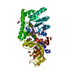
PDBj- Assembly
| Deposited unit | 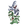
| ||||||||
|---|---|---|---|---|---|---|---|---|---|
| 1 |
| ||||||||
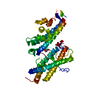
| Unit cell |
|
-Components
| #1: Protein | Mass: 23211.604 Da / Num. of mol.: 2 Source method: isolated from a genetically manipulated source Source: (gene. exp.) #2: Sugar | 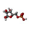  Type: D-saccharide, beta linking / Mass: 230.110 Da / Num. of mol.: 2 Type: D-saccharide, beta linking / Mass: 230.110 Da / Num. of mol.: 2Source method: isolated from a genetically manipulated source Formula: C5H11O8P #3: Water | ChemComp-HOH / |  Mass: 18.015 Da / Num. of mol.: 159 / Source method: isolated from a natural source / Formula: H2O Mass: 18.015 Da / Num. of mol.: 159 / Source method: isolated from a natural source / Formula: H2OHas protein modification | Y | |
|---|
-Experimental details
-Experiment
| Experiment | Method: X-RAY DIFFRACTION / Number of used crystals: 1 |
|---|
- Sample preparation
| Crystal | Density Matthews: 2.3 Å3/Da / Density % sol: 46 % |
|---|---|
| Crystal grow | pH: 8.5 Details: CRYSTALS WERE GROWN FROM A SOLUTION CONTAINING 3.5 MG/ML PROTEIN, 30-35 % PEG 4000 10 MM ARABINOSE-5-PHOSPHATE, 0.05M TRIS-HCL PH 8.4, 0.1 M MGCL2 |
-Data collection
| Diffraction | Mean temperature: 100 K |
|---|---|
| Diffraction source | Source: SYNCHROTRON / Site: ESRF  / Beamline: ID14-1 / Wavelength: 0.934 / Beamline: ID14-1 / Wavelength: 0.934 |
| Detector | Type: ADSC CCD / Detector: CCD / Date: Jul 13, 2002 / Details: GE(220),HORIZONTAL FOCUSING MULTILAYER |
| Radiation | Monochromator: DIAMOND (111), GE(220) / Protocol: SINGLE WAVELENGTH / Monochromatic (M) / Laue (L): M / Scattering type: x-ray |
| Radiation wavelength | Wavelength: 0.934 Å / Relative weight: 1 |
| Reflection | Resolution: 1.25→40 Å / Num. obs: 90067 / % possible obs: 81.8 % / Redundancy: 4.3 % / Rsym value: 0.095 / Net I/σ(I): 12.4 |
| Reflection shell | Resolution: 1.25→1.32 Å / Redundancy: 3.6 % / Mean I/σ(I) obs: 1.1 / Rsym value: 0.76 / % possible all: 40.1 |
- Processing
| Software |
| ||||||||||||||||||||||||||||||||||||||||||||||||||||||||||||||||||||||||||||||||||||||||||||||||||||||||||||||||||||||||||||||||||||||||||||||||||||||||||||||||||||||||||||||||||||||
|---|---|---|---|---|---|---|---|---|---|---|---|---|---|---|---|---|---|---|---|---|---|---|---|---|---|---|---|---|---|---|---|---|---|---|---|---|---|---|---|---|---|---|---|---|---|---|---|---|---|---|---|---|---|---|---|---|---|---|---|---|---|---|---|---|---|---|---|---|---|---|---|---|---|---|---|---|---|---|---|---|---|---|---|---|---|---|---|---|---|---|---|---|---|---|---|---|---|---|---|---|---|---|---|---|---|---|---|---|---|---|---|---|---|---|---|---|---|---|---|---|---|---|---|---|---|---|---|---|---|---|---|---|---|---|---|---|---|---|---|---|---|---|---|---|---|---|---|---|---|---|---|---|---|---|---|---|---|---|---|---|---|---|---|---|---|---|---|---|---|---|---|---|---|---|---|---|---|---|---|---|---|---|---|
| Refinement | Method to determine structure: MOLECULAR REPLACEMENT Starting model: PDB ENTRY 1KS2 Resolution: 1.25→40 Å / Cor.coef. Fo:Fc: 0.942 / Cor.coef. Fo:Fc free: 0.934 / SU B: 1.076 / SU ML: 0.046 / TLS residual ADP flag: LIKELY RESIDUAL / Cross valid method: THROUGHOUT / ESU R: 0.06 / ESU R Free: 0.06 / Stereochemistry target values: MAXIMUM LIKELIHOOD Details: ELECTRON DENSITY COULD NOT BE OBSERVED FOR RESIDUES 1-22, 32-46, 56-67, 98 AND 219 IN MOLECULE A AND RESIDUES 1,18-21, 98 AND 219 IN MOLECULE B. THESE RESIDUES WERE OMITTED AND ARE NOT PRESENT IN THE MODEL.
| ||||||||||||||||||||||||||||||||||||||||||||||||||||||||||||||||||||||||||||||||||||||||||||||||||||||||||||||||||||||||||||||||||||||||||||||||||||||||||||||||||||||||||||||||||||||
| Solvent computation | Ion probe radii: 0.8 Å / Shrinkage radii: 0.8 Å / VDW probe radii: 1.4 Å / Solvent model: BABINET MODEL WITH MASK | ||||||||||||||||||||||||||||||||||||||||||||||||||||||||||||||||||||||||||||||||||||||||||||||||||||||||||||||||||||||||||||||||||||||||||||||||||||||||||||||||||||||||||||||||||||||
| Displacement parameters | Biso mean: 10.26 Å2
| ||||||||||||||||||||||||||||||||||||||||||||||||||||||||||||||||||||||||||||||||||||||||||||||||||||||||||||||||||||||||||||||||||||||||||||||||||||||||||||||||||||||||||||||||||||||
| Refinement step | Cycle: LAST / Resolution: 1.25→40 Å
| ||||||||||||||||||||||||||||||||||||||||||||||||||||||||||||||||||||||||||||||||||||||||||||||||||||||||||||||||||||||||||||||||||||||||||||||||||||||||||||||||||||||||||||||||||||||
| Refine LS restraints |
|