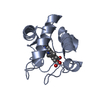
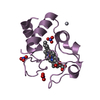
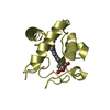
 Movie
Movie Controller


+ Open data

- Basic information
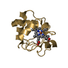
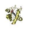
| Entry | Database: PDB / ID: 6duj | |||||||||
|---|---|---|---|---|---|---|---|---|---|---|
| Title | Crystal structure of A51V variant of Human Cytochrome c | |||||||||
 Components Components | Cytochrome c | |||||||||
 Keywords Keywords | APOPTOSIS / peroxidase activity / heme | |||||||||
| Function / homology |  Function and homology information Function and homology informationFormation of apoptosome / apoptosome / Release of apoptotic factors from the mitochondria / Respiratory electron transport / Activation of caspases through apoptosome-mediated cleavage / SMAC (DIABLO) binds to IAPs / SMAC(DIABLO)-mediated dissociation of IAP:caspase complexes / Regulation of the apoptosome activity / cellular respiration / mitochondrial electron transport, cytochrome c to oxygen ...Formation of apoptosome / apoptosome / Release of apoptotic factors from the mitochondria / Respiratory electron transport / Activation of caspases through apoptosome-mediated cleavage / SMAC (DIABLO) binds to IAPs / SMAC(DIABLO)-mediated dissociation of IAP:caspase complexes / Regulation of the apoptosome activity / cellular respiration / mitochondrial electron transport, cytochrome c to oxygen / execution phase of apoptosis / mitochondrial electron transport, ubiquinol to cytochrome c / Detoxification of Reactive Oxygen Species / Pyroptosis / intrinsic apoptotic signaling pathway / TP53 Regulates Metabolic Genes / apoptotic signaling pathway / Transcriptional activation of mitochondrial biogenesis / Cytoprotection by HMOX1 / mitochondrial intermembrane space / electron transfer activity / mitochondrial inner membrane / heme binding / mitochondrion / metal ion binding / nucleus / cytosol Similarity search - Function | |||||||||
| Biological species |  Homo sapiens (human) Homo sapiens (human) | |||||||||
| Method |  X-RAY DIFFRACTION / SYNCHROTRON / MOLECULAR REPLACEMENT / Resolution: 1.82202726164 Å X-RAY DIFFRACTION / SYNCHROTRON / MOLECULAR REPLACEMENT / Resolution: 1.82202726164 Å | |||||||||
 Authors Authors | Lei, H. / Bowler, B.E. | |||||||||
| Funding support |  United States, 2items United States, 2items
| |||||||||
 Citation Citation | Journal: J.Phys.Chem.B / Year: 2019 Title: Naturally Occurring A51V Variant of Human CytochromecDestabilizes the Native State and Enhances Peroxidase Activity. Authors: Lei, H. / Bowler, B.E. | |||||||||
| History |
|
- Structure visualization
| Structure viewer | Molecule: MolmilJmol/JSmol |
|---|
- Downloads & links
-Download
| PDBx/mmCIF format | 6duj.cif.gz | 116.8 KB | Display | PDBx/mmCIF format |
|---|---|---|---|---|
| PDB format | pdb6duj.ent.gz | 74.8 KB | Display | PDB format |
| PDBx/mmJSON format | 6duj.json.gz | Tree view | PDBx/mmJSON format | |
| Others |  Other downloads Other downloads |
-Validation report
| Arichive directory | https://data.pdbj.org/pub/pdb/validation_reports/du/6dujftp://data.pdbj.org/pub/pdb/validation_reports/du/6duj | HTTPS FTP |
|---|
-Related structure data
| Related structure data |  5ty3S S: Starting model for refinement |
|---|---|
| Similar structure data |


-Links
 PDBj
PDBj


- Assembly
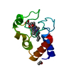
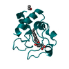
| Deposited unit | 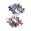
| ||||||||||||
|---|---|---|---|---|---|---|---|---|---|---|---|---|---|
| 1 | 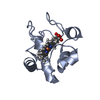
| ||||||||||||
| 2 | 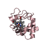
| ||||||||||||
| Unit cell |
| ||||||||||||
| Components on special symmetry positions |
|
-Components
| #1: Protein | Mass: 11668.636 Da / Num. of mol.: 2 / Mutation: A51V Source method: isolated from a genetically manipulated source Source: (gene. exp.) Homo sapiens (human) / Gene: CYCS, CYC / Production host:  #2: Chemical |   Mass: 618.503 Da / Num. of mol.: 2 / Source method: obtained synthetically / Formula: C34H34FeN4O4 Mass: 618.503 Da / Num. of mol.: 2 / Source method: obtained synthetically / Formula: C34H34FeN4O4#3: Water | ChemComp-HOH / |  Mass: 18.015 Da / Num. of mol.: 91 / Source method: isolated from a natural source / Formula: H2O Mass: 18.015 Da / Num. of mol.: 91 / Source method: isolated from a natural source / Formula: H2OHas protein modification | Y | |
|---|
-Experimental details
-Experiment
| Experiment | Method: X-RAY DIFFRACTION / Number of used crystals: 1 |
|---|
- Sample preparation
| Crystal | Density Matthews: 2.2 Å3/Da / Density % sol: 44.13 % |
|---|---|
| Crystal grow | Temperature: 293.15 K / Method: vapor diffusion, sitting drop / Details: 0.1 M sodium citrate pH 5.5, 40%(w/v) PEG 600 / PH range: 5.5-7.4 |
-Data collection
| Diffraction | Mean temperature: 100 K |
|---|---|
| Diffraction source | Source: SYNCHROTRON / Site: SSRL / Beamline: BL9-2 / Wavelength: 1.07 Å |
| Detector | Type: DECTRIS PILATUS 6M-F / Detector: PIXEL / Date: Mar 22, 2018 |
| Radiation | Protocol: SINGLE WAVELENGTH / Monochromatic (M) / Laue (L): M / Scattering type: x-ray |
| Radiation wavelength | Wavelength: 1.07 Å / Relative weight: 1 |
| Reflection | Resolution: 1.82→33.33 Å / Num. obs: 18770 / % possible obs: 98.7 % / Redundancy: 6.5 % / Biso Wilson estimate: 31.26 Å2 / Rmerge(I) obs: 0.106 / Net I/σ(I): 24.5 |
| Reflection shell | Resolution: 1.83→1.89 Å / Redundancy: 5.3 % / Rmerge(I) obs: 1.18 / Num. unique obs: 1878 / % possible all: 99 |
- Processing
| Software |
| ||||||||||||||||||||||||||||||||||||||||||||||||||||||||||||||||||||||||||||||||||||||||||||||||||
|---|---|---|---|---|---|---|---|---|---|---|---|---|---|---|---|---|---|---|---|---|---|---|---|---|---|---|---|---|---|---|---|---|---|---|---|---|---|---|---|---|---|---|---|---|---|---|---|---|---|---|---|---|---|---|---|---|---|---|---|---|---|---|---|---|---|---|---|---|---|---|---|---|---|---|---|---|---|---|---|---|---|---|---|---|---|---|---|---|---|---|---|---|---|---|---|---|---|---|---|
| Refinement | Method to determine structure: MOLECULAR REPLACEMENT Starting model: 5TY3 Resolution: 1.82202726164→33.3231225686 Å / Cross valid method: FREE R-VALUE / σ(F): 1.33795974971
| ||||||||||||||||||||||||||||||||||||||||||||||||||||||||||||||||||||||||||||||||||||||||||||||||||
| Displacement parameters | Biso mean: 35.2155113493 Å2 | ||||||||||||||||||||||||||||||||||||||||||||||||||||||||||||||||||||||||||||||||||||||||||||||||||
| Refinement step | Cycle: LAST / Resolution: 1.82202726164→33.3231225686 Å
| ||||||||||||||||||||||||||||||||||||||||||||||||||||||||||||||||||||||||||||||||||||||||||||||||||
| Refine LS restraints |
| ||||||||||||||||||||||||||||||||||||||||||||||||||||||||||||||||||||||||||||||||||||||||||||||||||
| LS refinement shell |
|
