 Movie
Movie Controller


+ Open data

- Basic information
| Entry | Database: PDB / ID: 6du8 | ||||||
|---|---|---|---|---|---|---|---|
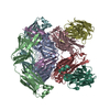
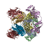
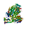
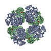
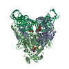
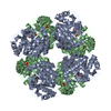
| Title | Human Polycsytin 2-l1 | ||||||
 Components Components | Polycystic kidney disease 2-like 1 protein | ||||||
 Keywords Keywords | MEMBRANE PROTEIN / polycystic 2-l1 / pc2l1 / pkd2l1 / TRPP2 | ||||||
| Function / homology |  Function and homology information Function and homology informationsour taste receptor activity / detection of chemical stimulus involved in sensory perception of sour taste / detection of chemical stimulus involved in sensory perception of taste / sensory perception of sour taste / response to water / osmolarity-sensing monoatomic cation channel activity / pH-gated monoatomic ion channel activity / calcium-activated potassium channel activity / muscle alpha-actinin binding / detection of mechanical stimulus ...sour taste receptor activity / detection of chemical stimulus involved in sensory perception of sour taste / detection of chemical stimulus involved in sensory perception of taste / sensory perception of sour taste / response to water / osmolarity-sensing monoatomic cation channel activity / pH-gated monoatomic ion channel activity / calcium-activated potassium channel activity / muscle alpha-actinin binding / detection of mechanical stimulus / non-motile cilium / : / calcium-activated cation channel activity / smoothened signaling pathway / ciliary membrane / sodium channel activity / alpha-actinin binding / monoatomic cation transmembrane transport / monoatomic cation transport / monoatomic cation channel activity / cellular response to acidic pH / cytoskeletal protein binding / calcium channel complex / potassium ion transmembrane transport / sodium ion transmembrane transport / calcium channel activity / actin cytoskeleton / cytoplasmic vesicle / protein homotetramerization / transmembrane transporter binding / signaling receptor complex / calcium ion binding / cell surface / endoplasmic reticulum / membrane / identical protein binding / plasma membrane / cytosol Similarity search - Function | ||||||
| Biological species |  Homo sapiens (human) Homo sapiens (human) | ||||||
| Method | ELECTRON MICROSCOPY / single particle reconstruction / cryo EM / Resolution: 3.11 Å | ||||||
 Authors Authors | Hulse, R.E. / Clapham, D.E. / Li, Z. / Huang, R.K. / Zhang, J. | ||||||
| Funding support |  United States, 1items United States, 1items
| ||||||
 Citation Citation | Journal: Elife / Year: 2018 Title: Cryo-EM structure of the polycystin 2-l1 ion channel. Authors: Raymond E Hulse / Zongli Li / Rick K Huang / Jin Zhang / David E Clapham /  Abstract: We report the near atomic resolution (3.3 Å) of the human polycystic kidney disease 2-like 1 (polycystin 2-l1) ion channel. Encoded by PKD2L1, polycystin 2-l1 is a calcium and monovalent cation- ...We report the near atomic resolution (3.3 Å) of the human polycystic kidney disease 2-like 1 (polycystin 2-l1) ion channel. Encoded by PKD2L1, polycystin 2-l1 is a calcium and monovalent cation-permeant ion channel in primary cilia and plasma membranes. The related primary cilium-specific polycystin-2 protein, encoded by PKD2, shares a high degree of sequence similarity, yet has distinct permeability characteristics. Here we show that these differences are reflected in the architecture of polycystin 2-l1. | ||||||
| History |
|
- Structure visualization
| Movie |
Movie viewer |
|---|---|
| Structure viewer | Molecule: MolmilJmol/JSmol |
- Downloads & links
-Download
| PDBx/mmCIF format | 6du8.cif.gz | 316.5 KB | Display | PDBx/mmCIF format |
|---|---|---|---|---|
| PDB format | pdb6du8.ent.gz | 246.3 KB | Display | PDB format |
| PDBx/mmJSON format | 6du8.json.gz | Tree view | PDBx/mmJSON format | |
| Others |  Other downloads Other downloads |
-Validation report
| Arichive directory | https://data.pdbj.org/pub/pdb/validation_reports/du/6du8ftp://data.pdbj.org/pub/pdb/validation_reports/du/6du8 | HTTPS FTP |
|---|
-Related structure data
| Related structure data |  8912MC M: map data used to model this data C: citing same article ( |
|---|---|
| Similar structure data |


-Links
 PDBj
PDBj
- Assembly
| Deposited unit | 
|
|---|---|
| 1 |
|
-Components
| #1: Protein | Mass: 92070.633 Da / Num. of mol.: 4 Source method: isolated from a genetically manipulated source Source: (gene. exp.) Homo sapiens (human) / Gene: PKD2L1, PKD2L, PKDL, TRPP3 / Plasmid: pEG / Cell (production host): HEK-293S GnTl / Cell line (production host): HEK293S / Production host: Homo sapiens (human) / References: UniProt: Q9P0L9#2: Sugar | ChemComp-NAG /   Type: D-saccharide, beta linking / Mass: 221.208 Da / Num. of mol.: 4 Type: D-saccharide, beta linking / Mass: 221.208 Da / Num. of mol.: 4Source method: isolated from a genetically manipulated source Formula: C8H15NO6 Has protein modification | Y | |
|---|
-Experimental details
-Experiment
| Experiment | Method: ELECTRON MICROSCOPY |
|---|---|
| EM experiment | Aggregation state: 3D ARRAY / 3D reconstruction method: single particle reconstruction |
- Sample preparation
| Component | Name: Human polycystic 2-l / Type: ORGANELLE OR CELLULAR COMPONENT / Details: Homotetrameric assembly of polycystic 2-l1 / Entity ID: #1 / Source: RECOMBINANT | |||||||||||||||||||||||||
|---|---|---|---|---|---|---|---|---|---|---|---|---|---|---|---|---|---|---|---|---|---|---|---|---|---|---|
| Molecular weight | Value: 0.547 MDa / Experimental value: NO | |||||||||||||||||||||||||
| Source (natural) | Organism: Homo sapiens (human) | |||||||||||||||||||||||||
| Source (recombinant) | Organism: Homo sapiens (human) / Plasmid: pEG | |||||||||||||||||||||||||
| Buffer solution | pH: 7.5 | |||||||||||||||||||||||||
| Buffer component |
| |||||||||||||||||||||||||
| Specimen | Conc.: 3.5 mg/ml / Embedding applied: NO / Shadowing applied: NO / Staining applied: NO / Vitrification applied: YES / Details: polycystic 2-l1 stabilized in amphipol PMAL-C8 | |||||||||||||||||||||||||
| Specimen support | Grid material: COPPER / Grid mesh size: 400 divisions/in. / Grid type: Quantifoil R1.2/1.3 | |||||||||||||||||||||||||
| Vitrification | Instrument: FEI VITROBOT MARK IV / Cryogen name: ETHANE / Humidity: 100 % / Chamber temperature: 277.15 K |
- Electron microscopy imaging
| Experimental equipment |  Model: Titan Krios / Image courtesy: FEI Company |
|---|---|
| Microscopy | Model: FEI TITAN KRIOS |
| Electron gun | Electron source:  FIELD EMISSION GUN / Accelerating voltage: 300 kV / Illumination mode: OTHER FIELD EMISSION GUN / Accelerating voltage: 300 kV / Illumination mode: OTHER |
| Electron lens | Mode: OTHER |
| Image recording | Electron dose: 60 e/Å2 / Film or detector model: GATAN K2 SUMMIT (4k x 4k) |
- Processing
| Software | Name: PHENIX / Version: 1.13_2998: / Classification: refinement |
|---|---|
| CTF correction | Type: PHASE FLIPPING AND AMPLITUDE CORRECTION |
| Symmetry | Point symmetry: C4 (4 fold cyclic) |
| 3D reconstruction | Resolution: 3.11 Å / Resolution method: FSC 0.143 CUT-OFF / Num. of particles: 119334 / Num. of class averages: 8 / Symmetry type: POINT |
