 Movie
Movie Controller


+ Open data

- Basic information
| Entry | Database: PDB / ID: 6dsi | |||||||||
|---|---|---|---|---|---|---|---|---|---|---|
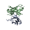
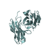
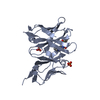
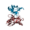
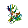
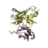
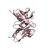
| Title | Anti recombinant prolactin receptor scFv | |||||||||
 Components Components | Anti-TN-C scFv | |||||||||
 Keywords Keywords | IMMUNE SYSTEM / anti prolactin receptor scFV | |||||||||
| Function / homology | Immunoglobulins / Immunoglobulin-like / Sandwich / Mainly Beta Function and homology information Function and homology information | |||||||||
| Biological species |  Homo sapiens (human) Homo sapiens (human) | |||||||||
| Method |  X-RAY DIFFRACTION / SYNCHROTRON / MOLECULAR REPLACEMENT / molecular replacement / Resolution: 2.49 Å X-RAY DIFFRACTION / SYNCHROTRON / MOLECULAR REPLACEMENT / molecular replacement / Resolution: 2.49 Å | |||||||||
 Authors Authors | Langley, D.B. / Rouet, R. / Christ, D. | |||||||||
| Funding support |  Australia, 2items Australia, 2items
| |||||||||
 Citation Citation | Journal: To Be Published Title: Anti recombinant prolactin receptor scFv Authors: Rouet, R. / Langley, D.B. / Christ, D. | |||||||||
| History |
|
- Structure visualization
| Structure viewer | Molecule: MolmilJmol/JSmol |
|---|
- Downloads & links
-Download
| PDBx/mmCIF format | 6dsi.cif.gz | 57.9 KB | Display | PDBx/mmCIF format |
|---|---|---|---|---|
| PDB format | pdb6dsi.ent.gz | 40.5 KB | Display | PDB format |
| PDBx/mmJSON format | 6dsi.json.gz | Tree view | PDBx/mmJSON format | |
| Others |  Other downloads Other downloads |
-Validation report
| Arichive directory | https://data.pdbj.org/pub/pdb/validation_reports/ds/6dsiftp://data.pdbj.org/pub/pdb/validation_reports/ds/6dsi | HTTPS FTP |
|---|
-Related structure data
| Related structure data |  6dn0S S: Starting model for refinement |
|---|---|
| Similar structure data |


-Links
 PDBj
PDBj

- Assembly
| Deposited unit | 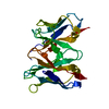
| ||||||||
|---|---|---|---|---|---|---|---|---|---|
| 1 |
| ||||||||
| Unit cell |
|
-Components
| #1: Antibody | Mass: 25560.133 Da / Num. of mol.: 1 Source method: isolated from a genetically manipulated source Source: (gene. exp.) Homo sapiens (human) / Production host:  |
|---|---|
| #2: Water | ChemComp-HOH /  Mass: 18.015 Da / Num. of mol.: 20 / Source method: isolated from a natural source / Formula: H2O Mass: 18.015 Da / Num. of mol.: 20 / Source method: isolated from a natural source / Formula: H2O |
| Has protein modification | Y |
-Experimental details
-Experiment
| Experiment | Method: X-RAY DIFFRACTION / Number of used crystals: 1 |
|---|
- Sample preparation
| Crystal | Density Matthews: 3.53 Å3/Da / Density % sol: 65.15 % |
|---|---|
| Crystal grow | Temperature: 293 K / Method: vapor diffusion, hanging drop / pH: 9 Details: Equal volumes (2 microlitres) of protein solution (5 mg/mL in 15 mM Tris (pH 7.5) and 50 mM NaCl) were combined with well solution (2% (v/v) Dioxane, 100 mM Bicine (pH 9.0) and 10% (w/v) PEG4000) |
-Data collection
| Diffraction | Mean temperature: 100 K |
|---|---|
| Diffraction source | Source: SYNCHROTRON / Site: Australian Synchrotron / Beamline: MX2 / Wavelength: 0.9537 Å |
| Detector | Type: ADSC QUANTUM 315r / Detector: CCD / Date: Nov 7, 2014 |
| Radiation | Protocol: SINGLE WAVELENGTH / Monochromatic (M) / Laue (L): M / Scattering type: x-ray |
| Radiation wavelength | Wavelength: 0.9537 Å / Relative weight: 1 |
| Reflection | Resolution: 2.49→37.8 Å / Num. obs: 12684 / % possible obs: 99.9 % / Redundancy: 6.6 % / CC1/2: 0.972 / Rmerge(I) obs: 0.113 / Rpim(I) all: 0.049 / Rrim(I) all: 0.123 / Net I/σ(I): 10.1 |
| Reflection shell | Resolution: 2.49→2.59 Å / Redundancy: 6.5 % / Rmerge(I) obs: 0.513 / Num. unique obs: 1397 / CC1/2: 0.905 / Rpim(I) all: 0.216 / Rrim(I) all: 0.557 / % possible all: 100 |
-Phasing
| Phasing | Method: molecular replacement | |||||||||
|---|---|---|---|---|---|---|---|---|---|---|
| Phasing MR | Model details: Phaser MODE: MR_AUTO
|
- Processing
| Software |
| ||||||||||||||||||||||||||||||||||||||||||||||||||||||||||||
|---|---|---|---|---|---|---|---|---|---|---|---|---|---|---|---|---|---|---|---|---|---|---|---|---|---|---|---|---|---|---|---|---|---|---|---|---|---|---|---|---|---|---|---|---|---|---|---|---|---|---|---|---|---|---|---|---|---|---|---|---|---|
| Refinement | Method to determine structure: MOLECULAR REPLACEMENT Starting model: 6dn0 Resolution: 2.49→36.86 Å / Cor.coef. Fo:Fc: 0.922 / Cor.coef. Fo:Fc free: 0.916 / SU B: 8.097 / SU ML: 0.175 / SU R Cruickshank DPI: 0.3087 / Cross valid method: THROUGHOUT / σ(F): 0 / ESU R: 0.309 / ESU R Free: 0.236 Details: HYDROGENS HAVE BEEN ADDED IN THE RIDING POSITIONS U VALUES : REFINED INDIVIDUALLY
| ||||||||||||||||||||||||||||||||||||||||||||||||||||||||||||
| Solvent computation | Ion probe radii: 0.8 Å / Shrinkage radii: 0.8 Å / VDW probe radii: 1.2 Å | ||||||||||||||||||||||||||||||||||||||||||||||||||||||||||||
| Displacement parameters | Biso max: 116.27 Å2 / Biso mean: 43.716 Å2 / Biso min: 23.03 Å2
| ||||||||||||||||||||||||||||||||||||||||||||||||||||||||||||
| Refinement step | Cycle: final / Resolution: 2.49→36.86 Å
| ||||||||||||||||||||||||||||||||||||||||||||||||||||||||||||
| Refine LS restraints |
| ||||||||||||||||||||||||||||||||||||||||||||||||||||||||||||
| LS refinement shell | Resolution: 2.488→2.552 Å / Rfactor Rfree error: 0 / Total num. of bins used: 20
|
