 Movie
Movie Controller

[English] 日本語
 Yorodumi
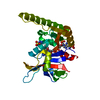
- PDB-6drs: Dihydrofolate Reductase (DHFR) of Aspergillus flavus in complex w... -
+ Open data

- Basic information
| Entry | Database: PDB / ID: 6drs | ||||||
|---|---|---|---|---|---|---|---|
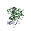
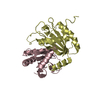
| Title | Dihydrofolate Reductase (DHFR) of Aspergillus flavus in complex with a small molecule inhibitor | ||||||
 Components Components | Dihydrofolate reductase, putative | ||||||
 Keywords Keywords | ANTIFUNGAL PROTEIN/Inhibitor / Antifolate / DHFR / ANTIFUNGAL PROTEIN / ANTIFUNGAL PROTEIN-Inhibitor complex | ||||||
| Function / homology |  Function and homology information Function and homology informationdihydrofolate metabolic process / dihydrofolate reductase / dihydrofolate reductase activity / folic acid metabolic process / tetrahydrofolate biosynthetic process / one-carbon metabolic process / NADP binding / mitochondrion Similarity search - Function | ||||||
| Biological species |  | ||||||
| Method |  X-RAY DIFFRACTION / MOLECULAR REPLACEMENT / Resolution: 1.997 Å X-RAY DIFFRACTION / MOLECULAR REPLACEMENT / Resolution: 1.997 Å | ||||||
 Authors Authors | Bensen, D.C. / Fortier, J.M. / Akers-Rodriguez, S. / Tari, L.W. | ||||||
 Citation Citation | Journal: To Be Published Title: Prospecting for broad-spectrum inhibitors of fungal dihydrofolate reductase using a structure guided approach. Authors: Bensen, D.C. / Fortier, J.M. / Akers-Rodriguez, S. / Tari, L.W. | ||||||
| History |
|
- Structure visualization
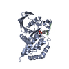
| Structure viewer | Molecule: MolmilJmol/JSmol |
|---|
- Downloads & links
-Download
| PDBx/mmCIF format | 6drs.cif.gz | 69.1 KB | Display | PDBx/mmCIF format |
|---|---|---|---|---|
| PDB format | pdb6drs.ent.gz | 48.4 KB | Display | PDB format |
| PDBx/mmJSON format | 6drs.json.gz | Tree view | PDBx/mmJSON format | |
| Others |  Other downloads Other downloads |
-Validation report
| Arichive directory | https://data.pdbj.org/pub/pdb/validation_reports/dr/6drsftp://data.pdbj.org/pub/pdb/validation_reports/dr/6drs | HTTPS FTP |
|---|
-Related structure data
| Related structure data |  6dtcC  4kyaS S: Starting model for refinement C: citing same article ( |
|---|---|
| Similar structure data |


-Links
 PDBj
PDBj
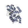
- Assembly
| Deposited unit | 
| ||||||||
|---|---|---|---|---|---|---|---|---|---|
| 1 |
| ||||||||
| Unit cell |
|
-Components
| #1: Protein | Mass: 27994.047 Da / Num. of mol.: 1 Source method: isolated from a genetically manipulated source Source: (gene. exp.) Strain: ATCC 200026 / FGSC A1120 / NRRL 3357 / JCM 12722 / SRRC 167 Gene: AFLA_046100 / Plasmid: pET28 / Production host:  | ||||||
|---|---|---|---|---|---|---|---|
| #2: Chemical | ChemComp-SO4 /   Mass: 96.063 Da / Num. of mol.: 5 / Source method: obtained synthetically / Formula: SO4 Mass: 96.063 Da / Num. of mol.: 5 / Source method: obtained synthetically / Formula: SO4#3: Chemical | ChemComp-H8A / | 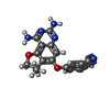  Mass: 333.344 Da / Num. of mol.: 1 / Source method: obtained synthetically / Formula: C18H15N5O2 Mass: 333.344 Da / Num. of mol.: 1 / Source method: obtained synthetically / Formula: C18H15N5O2#4: Chemical | ChemComp-NAP / |   Mass: 743.405 Da / Num. of mol.: 1 / Source method: obtained synthetically / Formula: C21H28N7O17P3 Mass: 743.405 Da / Num. of mol.: 1 / Source method: obtained synthetically / Formula: C21H28N7O17P3#5: Water | ChemComp-HOH / |  Mass: 18.015 Da / Num. of mol.: 160 / Source method: isolated from a natural source / Formula: H2O Mass: 18.015 Da / Num. of mol.: 160 / Source method: isolated from a natural source / Formula: H2O |
-Experimental details
-Experiment
| Experiment | Method: X-RAY DIFFRACTION / Number of used crystals: 1 |
|---|
- Sample preparation
| Crystal | Density Matthews: 3.95 Å3/Da / Density % sol: 68.88 % |
|---|---|
| Crystal grow | Temperature: 293 K / Method: evaporation Details: 0.1 M MES monohydrate pH 6.0, 2.4 M Ammonium sulfate |
-Data collection
| Diffraction | Mean temperature: 100 K | |||||||||||||||||||||||||||||||||||||||||||||||||||||||||||||||||||||||||||||||||||||||||||||||||||
|---|---|---|---|---|---|---|---|---|---|---|---|---|---|---|---|---|---|---|---|---|---|---|---|---|---|---|---|---|---|---|---|---|---|---|---|---|---|---|---|---|---|---|---|---|---|---|---|---|---|---|---|---|---|---|---|---|---|---|---|---|---|---|---|---|---|---|---|---|---|---|---|---|---|---|---|---|---|---|---|---|---|---|---|---|---|---|---|---|---|---|---|---|---|---|---|---|---|---|---|---|
| Diffraction source | Source: ROTATING ANODE / Type: RIGAKU RU200 / Wavelength: 1.5418 Å | |||||||||||||||||||||||||||||||||||||||||||||||||||||||||||||||||||||||||||||||||||||||||||||||||||
| Detector | Type: MAR scanner 345 mm plate / Detector: IMAGE PLATE / Date: Feb 23, 2015 | |||||||||||||||||||||||||||||||||||||||||||||||||||||||||||||||||||||||||||||||||||||||||||||||||||
| Radiation | Protocol: SINGLE WAVELENGTH / Monochromatic (M) / Laue (L): M / Scattering type: x-ray | |||||||||||||||||||||||||||||||||||||||||||||||||||||||||||||||||||||||||||||||||||||||||||||||||||
| Radiation wavelength | Wavelength: 1.5418 Å / Relative weight: 1 | |||||||||||||||||||||||||||||||||||||||||||||||||||||||||||||||||||||||||||||||||||||||||||||||||||
| Reflection | Resolution: 1.997→67.307 Å / Num. obs: 29252 / % possible obs: 99.9 % / Redundancy: 5.2 % / Rpim(I) all: 0.047 / Rrim(I) all: 0.11 / Rsym value: 0.099 / Net I/av σ(I): 6.7 / Net I/σ(I): 11.4 / Num. measured all: 152553 | |||||||||||||||||||||||||||||||||||||||||||||||||||||||||||||||||||||||||||||||||||||||||||||||||||
| Reflection shell | Diffraction-ID: 1
|
- Processing
| Software |
| |||||||||||||||||||||||||||||||||||||||||||||
|---|---|---|---|---|---|---|---|---|---|---|---|---|---|---|---|---|---|---|---|---|---|---|---|---|---|---|---|---|---|---|---|---|---|---|---|---|---|---|---|---|---|---|---|---|---|---|
| Refinement | Method to determine structure: MOLECULAR REPLACEMENT Starting model: 4KYA Resolution: 1.997→44.96 Å / Cor.coef. Fo:Fc: 0.931 / Cor.coef. Fo:Fc free: 0.895 / SU B: 4.713 / SU ML: 0.121 / Cross valid method: THROUGHOUT / σ(F): 0 / ESU R: 0.148 / ESU R Free: 0.145 / Details: U VALUES : REFINED INDIVIDUALLY
| |||||||||||||||||||||||||||||||||||||||||||||
| Solvent computation | Ion probe radii: 0.8 Å / Shrinkage radii: 0.8 Å / VDW probe radii: 1.2 Å | |||||||||||||||||||||||||||||||||||||||||||||
| Displacement parameters | Biso max: 120.55 Å2 / Biso mean: 26.04 Å2 / Biso min: 8.9 Å2
| |||||||||||||||||||||||||||||||||||||||||||||
| Refinement step | Cycle: final / Resolution: 1.997→44.96 Å
| |||||||||||||||||||||||||||||||||||||||||||||
| Refine LS restraints |
| |||||||||||||||||||||||||||||||||||||||||||||
| LS refinement shell | Resolution: 1.997→2.049 Å / Rfactor Rfree error: 0 / Total num. of bins used: 20
|
