 Movie
Movie Controller

[English] 日本語
 Yorodumi
- PDB-6drk: Structure of TRPM2 ion channel receptor by single particle electr... -
+ Open data

- Basic information
| Entry | Database: PDB / ID: 6drk | ||||||||||||||||||||||||||||||||||||||||||
|---|---|---|---|---|---|---|---|---|---|---|---|---|---|---|---|---|---|---|---|---|---|---|---|---|---|---|---|---|---|---|---|---|---|---|---|---|---|---|---|---|---|---|---|
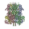
| Title | Structure of TRPM2 ion channel receptor by single particle electron cryo-microscopy, Apo state | ||||||||||||||||||||||||||||||||||||||||||
 Components Components | Transient receptor potential cation channel, subfamily M, member 2 | ||||||||||||||||||||||||||||||||||||||||||
 Keywords Keywords | TRANSPORT PROTEIN | ||||||||||||||||||||||||||||||||||||||||||
| Function / homology |  Function and homology information Function and homology informationTRP channels / Neutrophil degranulation / ADP-D-ribose binding / mono-ADP-D-ribose binding / ligand-gated calcium channel activity / ligand-gated monoatomic cation channel activity / monoatomic ion channel activity / calcium ion transmembrane transport / calcium channel activity / protein homotetramerization ...TRP channels / Neutrophil degranulation / ADP-D-ribose binding / mono-ADP-D-ribose binding / ligand-gated calcium channel activity / ligand-gated monoatomic cation channel activity / monoatomic ion channel activity / calcium ion transmembrane transport / calcium channel activity / protein homotetramerization / calcium ion binding / plasma membrane Similarity search - Function | ||||||||||||||||||||||||||||||||||||||||||
| Biological species |  | ||||||||||||||||||||||||||||||||||||||||||
| Method | ELECTRON MICROSCOPY / single particle reconstruction / cryo EM / Resolution: 3.8 Å | ||||||||||||||||||||||||||||||||||||||||||
 Authors Authors | Du, J. / Lu, W. / Huang, Y. / Winkler, P. / Sun, W. | ||||||||||||||||||||||||||||||||||||||||||
 Citation Citation | Journal: Nature / Year: 2018 Title: Architecture of the TRPM2 channel and its activation mechanism by ADP-ribose and calcium. Authors: Yihe Huang / Paige A Winkler / Weinan Sun / Wei Lü / Juan Du /  Abstract: Transient receptor potential melastatin 2 (TRPM2) is a calcium-permeable, non-selective cation channel that has an essential role in diverse physiological processes such as core body temperature ...Transient receptor potential melastatin 2 (TRPM2) is a calcium-permeable, non-selective cation channel that has an essential role in diverse physiological processes such as core body temperature regulation, immune response and apoptosis. TRPM2 is polymodal and can be activated by a wide range of stimuli, including temperature, oxidative stress and NAD-related metabolites such as ADP-ribose (ADPR). Its activation results in both Ca entry across the plasma membrane and Ca release from lysosomes, and has been linked to diseases such as ischaemia-reperfusion injury, bipolar disorder and Alzheimer's disease. Here we report the cryo-electron microscopy structures of the zebrafish TRPM2 in the apo resting (closed) state and in the ADPR/Ca-bound active (open) state, in which the characteristic NUDT9-H domains hang underneath the MHR1/2 domain. We identify an ADPR-binding site located in the bi-lobed structure of the MHR1/2 domain. Our results provide an insight into the mechanism of activation of the TRPM channel family and define a framework for the development of therapeutic agents to treat neurodegenerative diseases and temperature-related pathological conditions. | ||||||||||||||||||||||||||||||||||||||||||
| History |
|
- Structure visualization
| Movie |
Movie viewer |
|---|---|
| Structure viewer | Molecule: MolmilJmol/JSmol |
- Downloads & links
-Download
| PDBx/mmCIF format | 6drk.cif.gz | 801.6 KB | Display | PDBx/mmCIF format |
|---|---|---|---|---|
| PDB format | pdb6drk.ent.gz | 613.9 KB | Display | PDB format |
| PDBx/mmJSON format | 6drk.json.gz | Tree view | PDBx/mmJSON format | |
| Others |  Other downloads Other downloads |
-Validation report
| Arichive directory | https://data.pdbj.org/pub/pdb/validation_reports/dr/6drkftp://data.pdbj.org/pub/pdb/validation_reports/dr/6drk | HTTPS FTP |
|---|
-Related structure data
| Related structure data | 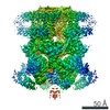 8901MC 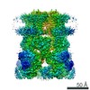 7999C 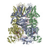 6drjC M: map data used to model this data C: citing same article ( |
|---|---|
| Similar structure data |

-Links
 PDBj
PDBj

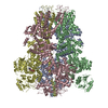
- Assembly
| Deposited unit | 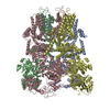
|
|---|---|
| 1 |
|
-Components
| #1: Protein | Mass: 168730.797 Da / Num. of mol.: 4 Source method: isolated from a genetically manipulated source Source: (gene. exp.)  Homo sapiens (human) / References: UniProt: A0A0R4IN04, UniProt: A0A0R4IMY7*PLUS Homo sapiens (human) / References: UniProt: A0A0R4IN04, UniProt: A0A0R4IMY7*PLUSHas protein modification | Y | |
|---|
-Experimental details
-Experiment
| Experiment | Method: ELECTRON MICROSCOPY |
|---|---|
| EM experiment | Aggregation state: PARTICLE / 3D reconstruction method: single particle reconstruction |
- Sample preparation
| Component | Name: Ion channel 1 / Type: COMPLEX / Entity ID: all / Source: RECOMBINANT |
|---|---|
| Molecular weight | Value: 0.5 MDa / Experimental value: YES |
| Source (natural) | Organism: |
| Source (recombinant) | Organism: Mammalia (mammals) |
| Buffer solution | pH: 8 |
| Specimen | Embedding applied: NO / Shadowing applied: NO / Staining applied: NO / Vitrification applied: YES |
| Vitrification | Cryogen name: ETHANE |
- Electron microscopy imaging
| Experimental equipment |  Model: Titan Krios / Image courtesy: FEI Company |
|---|---|
| Microscopy | Model: FEI TITAN KRIOS |
| Electron gun | Electron source:  FIELD EMISSION GUN / Accelerating voltage: 300 kV / Illumination mode: OTHER FIELD EMISSION GUN / Accelerating voltage: 300 kV / Illumination mode: OTHER |
| Electron lens | Mode: BRIGHT FIELD |
| Image recording | Electron dose: 45 e/Å2 / Film or detector model: GATAN K2 SUMMIT (4k x 4k) |
- Processing
| Software | Name: PHENIX / Version: dev_3084: / Classification: refinement |
|---|---|
| EM software | Name: PHENIX / Category: model refinement |
| CTF correction | Type: PHASE FLIPPING AND AMPLITUDE CORRECTION |
| 3D reconstruction | Resolution: 3.8 Å / Resolution method: FSC 0.143 CUT-OFF / Num. of particles: 183041 / Symmetry type: POINT |
