 Movie
Movie Controller

[English] 日本語
 Yorodumi
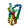
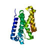
- PDB-6di2: Crystal structure of eukaryotic DNA primase large subunit iron-su... -
+ Open data

- Basic information
| Entry | Database: PDB / ID: 6di2 | ||||||
|---|---|---|---|---|---|---|---|
| Title | Crystal structure of eukaryotic DNA primase large subunit iron-sulfur cluster domain Y397L mutant | ||||||
 Components Components | DNA primase large subunit | ||||||
 Keywords Keywords | REPLICATION / DNA primase / p58 / iron-sulfur cluster | ||||||
| Function / homology |  Function and homology information Function and homology informationalpha DNA polymerase:primase complex / DNA replication, synthesis of primer / DNA replication initiation / 4 iron, 4 sulfur cluster binding / DNA binding / metal ion binding Similarity search - Function | ||||||
| Biological species |  | ||||||
| Method |  X-RAY DIFFRACTION / SYNCHROTRON / MOLECULAR REPLACEMENT / molecular replacement / Resolution: 1.32 Å X-RAY DIFFRACTION / SYNCHROTRON / MOLECULAR REPLACEMENT / molecular replacement / Resolution: 1.32 Å | ||||||
 Authors Authors | Salay, L.E. / Chazin, W.J. | ||||||
| Funding support |  United States, 1items United States, 1items
| ||||||
 Citation Citation | Journal: Proc. Natl. Acad. Sci. U.S.A. / Year: 2018 Title: Yeast require redox switching in DNA primase. Authors: O'Brien, E. / Salay, L.E. / Epum, E.A. / Friedman, K.L. / Chazin, W.J. / Barton, J.K. | ||||||
| History |
|
- Structure visualization
| Structure viewer | Molecule: MolmilJmol/JSmol |
|---|
- Downloads & links
-Download
| PDBx/mmCIF format | 6di2.cif.gz | 99.7 KB | Display | PDBx/mmCIF format |
|---|---|---|---|---|
| PDB format | pdb6di2.ent.gz | 73.2 KB | Display | PDB format |
| PDBx/mmJSON format | 6di2.json.gz | Tree view | PDBx/mmJSON format | |
| Others |  Other downloads Other downloads |
-Validation report
| Arichive directory | https://data.pdbj.org/pub/pdb/validation_reports/di/6di2ftp://data.pdbj.org/pub/pdb/validation_reports/di/6di2 | HTTPS FTP |
|---|
-Related structure data
| Related structure data |  6di6C  6dtvC  6dtzSC  6du0C C: citing same article ( S: Starting model for refinement |
|---|---|
| Similar structure data |
-Links
 PDBj
PDBj- Assembly
| Deposited unit | 
| ||||||||
|---|---|---|---|---|---|---|---|---|---|
| 1 |
| ||||||||
| Unit cell |
|
-Components
| #1: Protein | Mass: 23313.291 Da / Num. of mol.: 1 / Mutation: Y397L Source method: isolated from a genetically manipulated source Source: (gene. exp.) Strain: JAY291 / Gene: PRI2, C1Q_02024 / Production host:  References: UniProt: C7GP29, Transferases; Transferring phosphorus-containing groups; Nucleotidyltransferases | ||
|---|---|---|---|
| #2: Chemical | ChemComp-SF4 /   Mass: 351.640 Da / Num. of mol.: 1 / Source method: obtained synthetically / Formula: Fe4S4 Mass: 351.640 Da / Num. of mol.: 1 / Source method: obtained synthetically / Formula: Fe4S4 | ||
| #3: Chemical |   Mass: 118.174 Da / Num. of mol.: 2 / Source method: obtained synthetically / Formula: C6H14O2 / Comment: precipitant*YM Mass: 118.174 Da / Num. of mol.: 2 / Source method: obtained synthetically / Formula: C6H14O2 / Comment: precipitant*YM#4: Water | ChemComp-HOH / |  Mass: 18.015 Da / Num. of mol.: 201 / Source method: isolated from a natural source / Formula: H2O Mass: 18.015 Da / Num. of mol.: 201 / Source method: isolated from a natural source / Formula: H2O |
-Experimental details
-Experiment
| Experiment | Method: X-RAY DIFFRACTION / Number of used crystals: 1 |
|---|
- Sample preparation
| Crystal | Density Matthews: 2 Å3/Da / Density % sol: 38.42 % |
|---|---|
| Crystal grow | Temperature: 289 K / Method: vapor diffusion, hanging drop / pH: 8.5 / Details: MPD, TRIS-HCl |
-Data collection
| Diffraction | Mean temperature: 100 K |
|---|---|
| Diffraction source | Source: SYNCHROTRON / Site: APS / Beamline: 21-ID-D / Wavelength: 1.0787 Å |
| Detector | Type: DECTRIS EIGER X 9M / Detector: PIXEL / Date: Dec 10, 2016 |
| Radiation | Protocol: SINGLE WAVELENGTH / Monochromatic (M) / Laue (L): M / Scattering type: x-ray |
| Radiation wavelength | Wavelength: 1.0787 Å / Relative weight: 1 |
| Reflection | Resolution: 1.32→35 Å / Num. obs: 44491 / % possible obs: 99.2 % / Redundancy: 6.8 % / Biso Wilson estimate: 11.26 Å2 / Rmerge(I) obs: 0.066 / Rpim(I) all: 0.027 / Rrim(I) all: 0.072 / Χ2: 0.911 / Net I/σ(I): 11.7 / Num. measured all: 301921 |
| Reflection shell | Resolution: 1.32→1.37 Å / Redundancy: 6.4 % / Rmerge(I) obs: 0.184 / Num. unique obs: 4720 / CC1/2: 0.985 / Rpim(I) all: 0.027 / Rrim(I) all: 0.073 / Χ2: 1.049 / % possible all: 94.9 |
-Phasing
| Phasing | Method: molecular replacement | |||||||||
|---|---|---|---|---|---|---|---|---|---|---|
| Phasing MR | Model details: Phaser MODE: MR_AUTO
|
- Processing
| Software |
| ||||||||||||||||||||||||||||||||||||||||||||||||||||||||||||||||||||||||||||||||||||||||||||||||||||||||||||||||
|---|---|---|---|---|---|---|---|---|---|---|---|---|---|---|---|---|---|---|---|---|---|---|---|---|---|---|---|---|---|---|---|---|---|---|---|---|---|---|---|---|---|---|---|---|---|---|---|---|---|---|---|---|---|---|---|---|---|---|---|---|---|---|---|---|---|---|---|---|---|---|---|---|---|---|---|---|---|---|---|---|---|---|---|---|---|---|---|---|---|---|---|---|---|---|---|---|---|---|---|---|---|---|---|---|---|---|---|---|---|---|---|---|---|
| Refinement | Method to determine structure: MOLECULAR REPLACEMENT Starting model: 6DTZ Resolution: 1.32→33.62 Å / SU ML: 0.07 / Cross valid method: THROUGHOUT / σ(F): 1.37 / Phase error: 11.08
| ||||||||||||||||||||||||||||||||||||||||||||||||||||||||||||||||||||||||||||||||||||||||||||||||||||||||||||||||
| Solvent computation | Shrinkage radii: 0.9 Å / VDW probe radii: 1.11 Å | ||||||||||||||||||||||||||||||||||||||||||||||||||||||||||||||||||||||||||||||||||||||||||||||||||||||||||||||||
| Displacement parameters | Biso max: 52.02 Å2 / Biso mean: 17.5 Å2 / Biso min: 8 Å2 | ||||||||||||||||||||||||||||||||||||||||||||||||||||||||||||||||||||||||||||||||||||||||||||||||||||||||||||||||
| Refinement step | Cycle: final / Resolution: 1.32→33.62 Å
| ||||||||||||||||||||||||||||||||||||||||||||||||||||||||||||||||||||||||||||||||||||||||||||||||||||||||||||||||
| LS refinement shell | Refine-ID: X-RAY DIFFRACTION / Rfactor Rfree error: 0 / Total num. of bins used: 15
|
