 Movie
Movie Controller


+ Open data

- Basic information
| Entry | Database: PDB / ID: 6d87 | ||||||||||||
|---|---|---|---|---|---|---|---|---|---|---|---|---|---|
| Title | Structure of the Bovine p85alpha BH domain, R262T mutant | ||||||||||||
 Components Components | Phosphatidylinositol 3-kinase regulatory subunit alpha | ||||||||||||
 Keywords Keywords | SIGNALING PROTEIN / GAP protein | ||||||||||||
| Function / homology |  Function and homology information Function and homology informationRHOC GTPase cycle / PI3K events in ERBB4 signaling / Interleukin-7 signaling / GAB1 signalosome / PI3K events in ERBB2 signaling / MET activates PI3K/AKT signaling / CDC42 GTPase cycle / RHOJ GTPase cycle / RAC3 GTPase cycle / Erythropoietin activates Phosphoinositide-3-kinase (PI3K) ...RHOC GTPase cycle / PI3K events in ERBB4 signaling / Interleukin-7 signaling / GAB1 signalosome / PI3K events in ERBB2 signaling / MET activates PI3K/AKT signaling / CDC42 GTPase cycle / RHOJ GTPase cycle / RAC3 GTPase cycle / Erythropoietin activates Phosphoinositide-3-kinase (PI3K) / FLT3 Signaling / RND2 GTPase cycle / RND1 GTPase cycle / IRS-mediated signalling / GPVI-mediated activation cascade / Signaling by SCF-KIT / Downstream signal transduction / PI3K/AKT activation / Signaling by ALK / Role of phospholipids in phagocytosis / Tie2 Signaling / Role of LAT2/NTAL/LAB on calcium mobilization / CD28 dependent PI3K/Akt signaling / RAC2 GTPase cycle / Interleukin receptor SHC signaling / RND3 GTPase cycle / Co-stimulation by ICOS / PI-3K cascade:FGFR1 / PI-3K cascade:FGFR2 / PI-3K cascade:FGFR3 / PI-3K cascade:FGFR4 / Antigen activates B Cell Receptor (BCR) leading to generation of second messengers / PI3K Cascade / PIP3 activates AKT signaling / GP1b-IX-V activation signalling / RAF/MAP kinase cascade / PI5P, PP2A and IER3 Regulate PI3K/AKT Signaling / Synthesis of PIPs at the plasma membrane / RHOA GTPase cycle / RHOF GTPase cycle / DAP12 signaling / RHOU GTPase cycle / RHOV GTPase cycle / Regulation of signaling by CBL / Downstream TCR signaling / RHOG GTPase cycle / RET signaling / Interleukin-3, Interleukin-5 and GM-CSF signaling / VEGFA-VEGFR2 Pathway / phosphatidylinositol 3-kinase regulator activity / 1-phosphatidylinositol-3-kinase regulator activity / positive regulation of endoplasmic reticulum unfolded protein response / phosphatidylinositol 3-kinase activator activity / ErbB-3 class receptor binding / transmembrane receptor protein tyrosine kinase adaptor activity / phosphatidylinositol 3-kinase complex / phosphatidylinositol 3-kinase complex, class IA / Extra-nuclear estrogen signaling / G alpha (q) signalling events / phosphatidylinositol phosphate biosynthetic process / insulin receptor substrate binding / enzyme-substrate adaptor activity / phosphatidylinositol 3-kinase binding / insulin-like growth factor receptor binding / substrate adhesion-dependent cell spreading / insulin-like growth factor receptor signaling pathway / response to endoplasmic reticulum stress / intracellular glucose homeostasis / positive regulation of RNA splicing / positive regulation of D-glucose import across plasma membrane / GTPase activator activity / insulin receptor binding / positive regulation of protein localization to plasma membrane / phosphatidylinositol 3-kinase/protein kinase B signal transduction / positive regulation of protein import into nucleus / cellular response to insulin stimulus / insulin receptor signaling pathway / protein transport / protein stabilization / negative regulation of apoptotic process / positive regulation of transcription by RNA polymerase II / identical protein binding / nucleus Similarity search - Function | ||||||||||||
| Biological species |  | ||||||||||||
| Method |  X-RAY DIFFRACTION / SYNCHROTRON / MOLECULAR REPLACEMENT / Resolution: 2.7 Å X-RAY DIFFRACTION / SYNCHROTRON / MOLECULAR REPLACEMENT / Resolution: 2.7 Å | ||||||||||||
 Authors Authors | Moore, S.A. / Marshall, J.D. / Anderson, D.H. | ||||||||||||
| Funding support |  Canada, 3items Canada, 3items
| ||||||||||||
 Citation Citation | Journal: Sci Rep / Year: 2018 Title: Patient-derived mutations within the N-terminal domains of p85 alpha impact PTEN or Rab5 binding and regulation. Authors: Mellor, P. / Marshall, J.D.S. / Ruan, X. / Whitecross, D.E. / Ross, R.L. / Knowles, M.A. / Moore, S.A. / Anderson, D.H. | ||||||||||||
| History |
|
- Structure visualization
| Structure viewer | Molecule: MolmilJmol/JSmol |
|---|
- Downloads & links
-Download
| PDBx/mmCIF format | 6d87.cif.gz | 146.3 KB | Display | PDBx/mmCIF format |
|---|---|---|---|---|
| PDB format | pdb6d87.ent.gz | 116.3 KB | Display | PDB format |
| PDBx/mmJSON format | 6d87.json.gz | Tree view | PDBx/mmJSON format | |
| Others |  Other downloads Other downloads |
-Validation report
| Arichive directory | https://data.pdbj.org/pub/pdb/validation_reports/d8/6d87ftp://data.pdbj.org/pub/pdb/validation_reports/d8/6d87 | HTTPS FTP |
|---|
-Related structure data
| Related structure data |  6d81C  6d82C 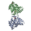 6d85C  6d86C  1pbwS S: Starting model for refinement C: citing same article ( |
|---|---|
| Similar structure data |


-Links
 PDBj
PDBj


- Assembly
| Deposited unit | 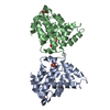
| ||||||||
|---|---|---|---|---|---|---|---|---|---|
| 1 |
| ||||||||
| Unit cell |
|
-Components
| #1: Protein | Mass: 21853.057 Da / Num. of mol.: 2 / Mutation: R262T Source method: isolated from a genetically manipulated source Source: (gene. exp.) Details (production host): Glutathione-S-Transferase fusion protein Production host:  #2: Chemical |   Mass: 96.063 Da / Num. of mol.: 3 / Source method: obtained synthetically / Formula: SO4 Mass: 96.063 Da / Num. of mol.: 3 / Source method: obtained synthetically / Formula: SO4#3: Water | ChemComp-HOH / |  Mass: 18.015 Da / Num. of mol.: 85 / Source method: isolated from a natural source / Formula: H2O Mass: 18.015 Da / Num. of mol.: 85 / Source method: isolated from a natural source / Formula: H2OHas protein modification | Y | |
|---|
-Experimental details
-Experiment
| Experiment | Method: X-RAY DIFFRACTION / Number of used crystals: 1 |
|---|
- Sample preparation
| Crystal | Density Matthews: 4.19 Å3/Da / Density % sol: 70.67 % / Description: Prism |
|---|---|
| Crystal grow | Temperature: 293 K / Method: vapor diffusion, hanging drop / pH: 6 / Details: 1.5 M Li2SO4, 100 mM Na Cacodylate, pH 6.0 |
-Data collection
| Diffraction | Mean temperature: 80 K |
|---|---|
| Diffraction source | Source: SYNCHROTRON / Site: CLSI / Beamline: 08B1-1 / Wavelength: 0.987 Å |
| Detector | Type: RAYONIX MX300HE / Detector: CCD / Date: Oct 25, 2015 / Details: Toroidal focusing mirrors |
| Radiation | Monochromator: Double crystal / Protocol: SINGLE WAVELENGTH / Monochromatic (M) / Laue (L): M / Scattering type: x-ray |
| Radiation wavelength | Wavelength: 0.987 Å / Relative weight: 1 |
| Reflection | Resolution: 2.7→35.8 Å / Num. obs: 20681 / % possible obs: 99.3 % / Observed criterion σ(F): 1 / Redundancy: 7.4 % / Biso Wilson estimate: 40.9 Å2 / Rmerge(I) obs: 0.089 / Net I/σ(I): 23.3 |
| Reflection shell | Resolution: 2.7→2.75 Å / Redundancy: 7.5 % / Rmerge(I) obs: 0.501 / Num. unique obs: 1024 / % possible all: 100 |
- Processing
| Software |
| |||||||||||||||||||||||||||||||||||||||||||||||||
|---|---|---|---|---|---|---|---|---|---|---|---|---|---|---|---|---|---|---|---|---|---|---|---|---|---|---|---|---|---|---|---|---|---|---|---|---|---|---|---|---|---|---|---|---|---|---|---|---|---|---|
| Refinement | Method to determine structure: MOLECULAR REPLACEMENT Starting model: 1PBW Resolution: 2.7→35.795 Å / SU ML: 0.31 / Cross valid method: FREE R-VALUE / σ(F): 1.35 / Phase error: 22.92 / Details: Standard ML refinement
| |||||||||||||||||||||||||||||||||||||||||||||||||
| Solvent computation | Shrinkage radii: 0.9 Å / VDW probe radii: 1.11 Å | |||||||||||||||||||||||||||||||||||||||||||||||||
| Refinement step | Cycle: LAST / Resolution: 2.7→35.795 Å
| |||||||||||||||||||||||||||||||||||||||||||||||||
| Refine LS restraints |
| |||||||||||||||||||||||||||||||||||||||||||||||||
| LS refinement shell |
|
