 Movie
Movie Controller

[English] 日本語
 Yorodumi
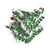
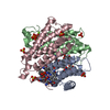
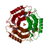
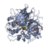
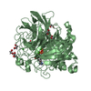
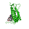
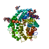
- PDB-6d5k: Structure of Human ATP:Cobalamin Adenosyltransferase bound to ATP... -
+ Open data

- Basic information
| Entry | Database: PDB / ID: 6d5k | ||||||
|---|---|---|---|---|---|---|---|
| Title | Structure of Human ATP:Cobalamin Adenosyltransferase bound to ATP, and Adenosylcobalamin | ||||||
 Components Components | Cob(I)yrinic acid a,c-diamide adenosyltransferase, mitochondrial | ||||||
 Keywords Keywords | TRANSFERASE / B12 / metabolism / adenosyltransferase | ||||||
| Function / homology |  Function and homology information Function and homology informationDefective MMAB causes MMA, cblB type / Cobalamin (Cbl) metabolism / cobalamin metabolic process / corrinoid adenosyltransferase activity / Transferases; Transferring alkyl or aryl groups, other than methyl groups / transferase activity, transferring alkyl or aryl (other than methyl) groups / cobalamin binding / mitochondrial matrix / mitochondrion / ATP binding Similarity search - Function | ||||||
| Biological species |  Homo sapiens (human) Homo sapiens (human) | ||||||
| Method |  X-RAY DIFFRACTION / SYNCHROTRON / MOLECULAR REPLACEMENT / molecular replacement / Resolution: 2.85 Å X-RAY DIFFRACTION / SYNCHROTRON / MOLECULAR REPLACEMENT / molecular replacement / Resolution: 2.85 Å | ||||||
 Authors Authors | Dodge, G.J. / Campanello, G. / Smith, J.L. / Banerjee, R. | ||||||
| Funding support |  United States, 1items United States, 1items
| ||||||
 Citation Citation | Journal: J. Am. Chem. Soc. / Year: 2018 Title: Sacrificial Cobalt-Carbon Bond Homolysis in Coenzyme B12as a Cofactor Conservation Strategy. Authors: Campanello, G.C. / Ruetz, M. / Dodge, G.J. / Gouda, H. / Gupta, A. / Twahir, U.T. / Killian, M.M. / Watkins, D. / Rosenblatt, D.S. / Brunold, T.C. / Warncke, K. / Smith, J.L. / Banerjee, R. | ||||||
| History |
|
- Structure visualization
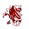
| Structure viewer | Molecule: MolmilJmol/JSmol |
|---|
- Downloads & links
-Download
| PDBx/mmCIF format | 6d5k.cif.gz | 126.6 KB | Display | PDBx/mmCIF format |
|---|---|---|---|---|
| PDB format | pdb6d5k.ent.gz | 95.6 KB | Display | PDB format |
| PDBx/mmJSON format | 6d5k.json.gz | Tree view | PDBx/mmJSON format | |
| Others |  Other downloads Other downloads |
-Validation report
| Arichive directory | https://data.pdbj.org/pub/pdb/validation_reports/d5/6d5kftp://data.pdbj.org/pub/pdb/validation_reports/d5/6d5k | HTTPS FTP |
|---|
-Related structure data
| Related structure data |  6d5xC  2idxS S: Starting model for refinement C: citing same article ( |
|---|---|
| Similar structure data |
-Links
 PDBj
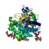
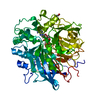
PDBj- Assembly
| Deposited unit | 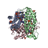
| |||||||||||||||||||||||||||||||||||||||||||||||||||||
|---|---|---|---|---|---|---|---|---|---|---|---|---|---|---|---|---|---|---|---|---|---|---|---|---|---|---|---|---|---|---|---|---|---|---|---|---|---|---|---|---|---|---|---|---|---|---|---|---|---|---|---|---|---|---|
| 1 |
| |||||||||||||||||||||||||||||||||||||||||||||||||||||
| 2 | 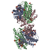
| |||||||||||||||||||||||||||||||||||||||||||||||||||||
| Unit cell |
| |||||||||||||||||||||||||||||||||||||||||||||||||||||
| Noncrystallographic symmetry (NCS) | NCS domain:
NCS domain segments: Ens-ID: 1
|
-Components
-Protein , 1 types, 3 molecules ABC
| #1: Protein | Mass: 21720.693 Da / Num. of mol.: 3 Source method: isolated from a genetically manipulated source Source: (gene. exp.) Homo sapiens (human) / Gene: MMAB / Production host:  |
|---|
-Non-polymers , 7 types, 32 molecules 


| #2: Chemical |  Mass: 251.242 Da / Num. of mol.: 2 / Source method: obtained synthetically / Formula: C10H13N5O3 / Feature type: SUBJECT OF INVESTIGATION Mass: 251.242 Da / Num. of mol.: 2 / Source method: obtained synthetically / Formula: C10H13N5O3 / Feature type: SUBJECT OF INVESTIGATION#3: Chemical |  Mass: 1330.356 Da / Num. of mol.: 2 / Source method: obtained synthetically / Formula: C62H89CoN13O14P / Feature type: SUBJECT OF INVESTIGATION Mass: 1330.356 Da / Num. of mol.: 2 / Source method: obtained synthetically / Formula: C62H89CoN13O14P / Feature type: SUBJECT OF INVESTIGATION#4: Chemical | ChemComp-SO4 /  Mass: 96.063 Da / Num. of mol.: 6 / Source method: obtained synthetically / Formula: SO4 Mass: 96.063 Da / Num. of mol.: 6 / Source method: obtained synthetically / Formula: SO4#5: Chemical | ChemComp-ATP / |  Mass: 507.181 Da / Num. of mol.: 1 Mass: 507.181 Da / Num. of mol.: 1Source method: isolated from a genetically manipulated source Formula: C10H16N5O13P3 / Feature type: SUBJECT OF INVESTIGATION / Comment: ATP, energy-carrying molecule*YM #6: Chemical |  Mass: 24.305 Da / Num. of mol.: 2 / Source method: obtained synthetically / Formula: Mg / Feature type: SUBJECT OF INVESTIGATION Mass: 24.305 Da / Num. of mol.: 2 / Source method: obtained synthetically / Formula: Mg / Feature type: SUBJECT OF INVESTIGATION#7: Chemical | ChemComp-EPE / |  Mass: 238.305 Da / Num. of mol.: 1 Mass: 238.305 Da / Num. of mol.: 1Source method: isolated from a genetically manipulated source Formula: C8H18N2O4S / Comment: pH buffer*YM #8: Water | ChemComp-HOH / | Mass: 18.015 Da / Num. of mol.: 18 / Source method: isolated from a natural source / Formula: H2O |
|---|
-Experimental details
-Experiment
| Experiment | Method: X-RAY DIFFRACTION / Number of used crystals: 1 |
|---|
- Sample preparation
| Crystal | Density Matthews: 3.29 Å3/Da / Density % sol: 62.65 % |
|---|---|
| Crystal grow | Temperature: 293 K / Method: vapor diffusion, sitting drop / pH: 7.5 / Details: 19% PEG 3350, 0.2 M MgSO4, 10 % glycerol |
-Data collection
| Diffraction | Mean temperature: 298 K | ||||||||||||||||||||||||
|---|---|---|---|---|---|---|---|---|---|---|---|---|---|---|---|---|---|---|---|---|---|---|---|---|---|
| Diffraction source | Source: SYNCHROTRON / Site: APS / Beamline: 23-ID-B / Wavelength: 1.5498 Å | ||||||||||||||||||||||||
| Detector | Type: DECTRIS EIGER X 16M / Detector: PIXEL / Date: Jun 8, 2017 | ||||||||||||||||||||||||
| Radiation | Monochromator: double crystal / Protocol: SINGLE WAVELENGTH / Monochromatic (M) / Laue (L): M / Scattering type: x-ray | ||||||||||||||||||||||||
| Radiation wavelength | Wavelength: 1.5498 Å / Relative weight: 1 | ||||||||||||||||||||||||
| Reflection | Resolution: 2.68→48.61 Å / Num. obs: 24504 / % possible obs: 99.9 % / Redundancy: 9.9 % / CC1/2: 0.997 / Rmerge(I) obs: 0.22 / Rpim(I) all: 0.074 / Rrim(I) all: 0.232 / Net I/σ(I): 7.6 / Num. measured all: 242325 / Scaling rejects: 1 | ||||||||||||||||||||||||
| Reflection shell | Diffraction-ID: 1
|
-Phasing
| Phasing | Method: molecular replacement | |||||||||
|---|---|---|---|---|---|---|---|---|---|---|
| Phasing MR |
|
- Processing
| Software |
| ||||||||||||||||||||||||
|---|---|---|---|---|---|---|---|---|---|---|---|---|---|---|---|---|---|---|---|---|---|---|---|---|---|
| Refinement | Method to determine structure: MOLECULAR REPLACEMENT Starting model: 2idx Resolution: 2.85→48.607 Å / SU ML: 0.48 / Cross valid method: THROUGHOUT / σ(F): 1.35 / Phase error: 26.56
| ||||||||||||||||||||||||
| Solvent computation | Shrinkage radii: 0.9 Å / VDW probe radii: 1.11 Å | ||||||||||||||||||||||||
| Displacement parameters | Biso max: 203.75 Å2 / Biso mean: 83.9279 Å2 / Biso min: 46.04 Å2 | ||||||||||||||||||||||||
| Refinement step | Cycle: final / Resolution: 2.85→48.607 Å
| ||||||||||||||||||||||||
| Refine LS restraints NCS |
|
