Resolution: 1.79→35.8 Å / Cor.coef. Fo:Fc: 0.8619 / Cor.coef. Fo:Fc free: 0.7979 / SU R Cruickshank DPI: 0.275 / Cross valid method: THROUGHOUT / σ(F): 0 / SU R Blow DPI: 0.189 / SU Rfree Blow DPI: 0.17 / SU Rfree Cruickshank DPI: 0.168 Details: IDEAL-DIST CONTACT TERM CONTACT SETUP RESIDUE TYPES WITHOUT CCP4 ATOM TYPE IN LIBRARY=FE CA. NUMBER OF ATOMS WITH PROPER CCP4 ATOM TYPE=9438. NUMBER WITH APPROX DEFAULT CCP4 ATOM TYPE=0. ...Details: IDEAL-DIST CONTACT TERM CONTACT SETUP RESIDUE TYPES WITHOUT CCP4 ATOM TYPE IN LIBRARY=FE CA. NUMBER OF ATOMS WITH PROPER CCP4 ATOM TYPE=9438. NUMBER WITH APPROX DEFAULT CCP4 ATOM TYPE=0. NUMBER TREATED BY BAD NON-BONDED CONTACTS=5.
Rfactor
Num. reflection
% reflection
Selection details
Rfree
0.2866
2614
5.06 %
RANDOM
Rwork
0.2428
-
-
-
obs
0.2451
51706
87.25 %
-
Displacement parameters
Biso mean: 18.48 Å2
Baniso -1
Baniso -2
Baniso -3
1-
-1.8853 Å2
0 Å2
0 Å2
2-
-
1.2594 Å2
0 Å2
3-
-
-
0.6259 Å2
Refine analyze
Luzzati coordinate error obs: 0.439 Å
Refinement step
Cycle: LAST / Resolution: 1.79→35.8 Å
Protein
Nucleic acid
Ligand
Solvent
Total
Num. atoms
4469
0
45
524
5038
Refine LS restraints
Refine-ID
Type
Dev ideal
Number
Restraint function
Weight
X-RAY DIFFRACTION
t_bond_d
0.01
9025
HARMONIC
2
X-RAY DIFFRACTION
t_angle_deg
1.11
16231
HARMONIC
2
X-RAY DIFFRACTION
t_dihedral_angle_d
1914
SINUSOIDAL
2
X-RAY DIFFRACTION
t_incorr_chiral_ct
X-RAY DIFFRACTION
t_pseud_angle
X-RAY DIFFRACTION
t_trig_c_planes
133
HARMONIC
2
X-RAY DIFFRACTION
t_gen_planes
1434
HARMONIC
5
X-RAY DIFFRACTION
t_it
9025
HARMONIC
20
X-RAY DIFFRACTION
t_nbd
0
SEMIHARMONIC
5
X-RAY DIFFRACTION
t_omega_torsion
3.8
X-RAY DIFFRACTION
t_other_torsion
14.25
X-RAY DIFFRACTION
t_improper_torsion
X-RAY DIFFRACTION
t_chiral_improper_torsion
582
SEMIHARMONIC
5
X-RAY DIFFRACTION
t_sum_occupancies
X-RAY DIFFRACTION
t_utility_distance
X-RAY DIFFRACTION
t_utility_angle
X-RAY DIFFRACTION
t_utility_torsion
X-RAY DIFFRACTION
t_ideal_dist_contact
10099
SEMIHARMONIC
4
LS refinement shell
Resolution: 1.79→1.84 Å / Total num. of bins used: 20
Rfactor
Num. reflection
% reflection
Rfree
0.2509
205
4.97 %
Rwork
0.2189
3919
-
all
0.2206
4124
-
obs
-
-
87.25 %
+
About Yorodumi
-
News
-
Feb 9, 2022. New format data for meta-information of EMDB entries
New format data for meta-information of EMDB entries
Version 3 of the EMDB header file is now the official format.
The previous official version 1.9 will be removed from the archive.
In the structure databanks used in Yorodumi, some data are registered as the other names, "COVID-19 virus" and "2019-nCoV". Here are the details of the virus and the list of structure data.
Jan 31, 2019. EMDB accession codes are about to change! (news from PDBe EMDB page)
EMDB accession codes are about to change! (news from PDBe EMDB page)
The allocation of 4 digits for EMDB accession codes will soon come to an end. Whilst these codes will remain in use, new EMDB accession codes will include an additional digit and will expand incrementally as the available range of codes is exhausted. The current 4-digit format prefixed with “EMD-” (i.e. EMD-XXXX) will advance to a 5-digit format (i.e. EMD-XXXXX), and so on. It is currently estimated that the 4-digit codes will be depleted around Spring 2019, at which point the 5-digit format will come into force.
The EM Navigator/Yorodumi systems omit the EMD- prefix.
Related info.:Q: What is EMD? / ID/Accession-code notation in Yorodumi/EM Navigator
Yorodumi is a browser for structure data from EMDB, PDB, SASBDB, etc.
This page is also the successor to EM Navigator detail page, and also detail information page/front-end page for Omokage search.
The word "yorodu" (or yorozu) is an old Japanese word meaning "ten thousand". "mi" (miru) is to see.
Related info.:EMDB / PDB / SASBDB / Comparison of 3 databanks / Yorodumi Search / Aug 31, 2016. New EM Navigator & Yorodumi / Yorodumi Papers / Jmol/JSmol / Function and homology information / Changes in new EM Navigator and Yorodumi
 Movie
Movie Controller

 Yorodumi
 Open data
 Basic information
 Components
 Keywords
 Function and homology information
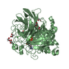
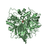
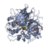
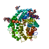
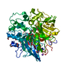
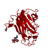
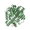
 PSEUDOMONAS FLUORESCENS (bacteria)
 X-RAY DIFFRACTION /
  Authors
 Citation
 Structure visualization
 Downloads & links
 Other downloads
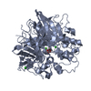


 PDBj
PDBj


 Assembly
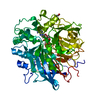


 Mass: 62.068 Da / Num. of mol.: 2 / Source method: obtained synthetically / Formula: C2H6O2
 Mass: 505.208 Da / Num. of mol.: 1 / Source method: obtained synthetically / Formula: C11H18N5O12P3 / Comment: AMP-PCP, energy-carrying molecule analogue*YM
 Mass: 40.078 Da / Num. of mol.: 3 / Source method: obtained synthetically / Formula: Ca
 Mass: 127.689 Da / Num. of mol.: 1 / Source method: obtained synthetically / Formula: Fe2O
 Sample preparation
 / Beamline: ID29 / Wavelength: 0.97932
  Processing