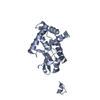
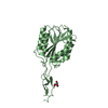
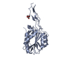
Mass: 15414.208 Da / Num. of mol.: 1 Source method: isolated from a genetically manipulated source Details: A gift from the lab of Dr. Roger C. Levesque Source: (gene. exp.) Pseudomonas aeruginosa LESB58 (bacteria) Gene: PAMH19_0675 / Plasmid: pMCSG11 Details (production host): pMCSG11 with fragement of PALES_06171 (R1-NTF) clones in frame into LIC site Production host: Escherichia coli BL21(DE3) (bacteria) / References: UniProt: A0A0A8RA06
Mass: 18.015 Da / Num. of mol.: 153 / Source method: isolated from a natural source / Formula: H2O
-
Experimental details
-
Experiment
Experiment
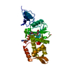
Method: X-RAY DIFFRACTION / Number of used crystals: 1
-
Sample preparation
Crystal
Density Matthews: 2.88 Å3/Da / Density % sol: 57.35 % / Description: Hexagonal prisms.
Crystal grow
Temperature: 291 K / Method: vapor diffusion, hanging drop Details: Crystals were grown at a 1:1 protein (5 mg/mL) to precipitant (100mM Bicine pH 9.5, 20% PEG2k, 1mM CuSO4) ratio
-
Data collection
Diffraction
Mean temperature: 120 K
Diffraction source
Source: SYNCHROTRON / Site: ALS / Beamline: 5.0.2 / Wavelength: 0.987 Å
In the structure databanks used in Yorodumi, some data are registered as the other names, "COVID-19 virus" and "2019-nCoV". Here are the details of the virus and the list of structure data.
Jan 31, 2019. EMDB accession codes are about to change! (news from PDBe EMDB page)
EMDB accession codes are about to change! (news from PDBe EMDB page)
The allocation of 4 digits for EMDB accession codes will soon come to an end. Whilst these codes will remain in use, new EMDB accession codes will include an additional digit and will expand incrementally as the available range of codes is exhausted. The current 4-digit format prefixed with “EMD-” (i.e. EMD-XXXX) will advance to a 5-digit format (i.e. EMD-XXXXX), and so on. It is currently estimated that the 4-digit codes will be depleted around Spring 2019, at which point the 5-digit format will come into force.
The EM Navigator/Yorodumi systems omit the EMD- prefix.
Related info.:Q: What is EMD? / ID/Accession-code notation in Yorodumi/EM Navigator
Yorodumi is a browser for structure data from EMDB, PDB, SASBDB, etc.
This page is also the successor to EM Navigator detail page, and also detail information page/front-end page for Omokage search.
The word "yorodu" (or yorozu) is an old Japanese word meaning "ten thousand". "mi" (miru) is to see.
Related info.:EMDB / PDB / SASBDB / Comparison of 3 databanks / Yorodumi Search / Aug 31, 2016. New EM Navigator & Yorodumi / Yorodumi Papers / Jmol/JSmol / Function and homology information / Changes in new EM Navigator and Yorodumi
 Movie
Movie Controller

 Yorodumi
 Open data
 Basic information
 Components
 Keywords
 Function and homology information
 Pseudomonas aeruginosa LESB58 (bacteria)
 X-RAY DIFFRACTION /
  Authors
 United States, 1items
  Citation
 Structure visualization
 Downloads & links
 Other downloads

 PDBj
PDBj Assembly

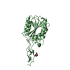
 Mass: 24.305 Da / Num. of mol.: 1 / Source method: obtained synthetically / Formula: Mg / Feature type: SUBJECT OF INVESTIGATION
 Mass: 92.094 Da / Num. of mol.: 1 / Source method: obtained synthetically / Formula: C3H8O3 / Feature type: SUBJECT OF INVESTIGATION
 Mass: 18.015 Da / Num. of mol.: 153 / Source method: isolated from a natural source / Formula: H2O
 Sample preparation
 Processing