 Movie
Movie Controller


+ Open data

- Basic information
| Entry | Database: PDB / ID: 6cv9 | |||||||||
|---|---|---|---|---|---|---|---|---|---|---|
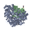
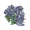
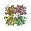
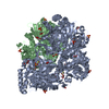
| Title | Cytoplasmic domain of mTRPC6 | |||||||||
 Components Components | Short transient receptor potential channel 6 | |||||||||
 Keywords Keywords | TRANSPORT PROTEIN / Ion Channel / Membrane Protein / TRP Channel / TRPC6 / focal segmental glomerulosclerosis | |||||||||
| Function / homology |  Function and homology information Function and homology informationpositive regulation of ion transmembrane transporter activity / Effects of PIP2 hydrolysis / Elevation of cytosolic Ca2+ levels / slit diaphragm / negative regulation of dendrite morphogenesis / store-operated calcium channel activity / TRP channels / cation channel complex / positive regulation of calcium ion transport / inositol 1,4,5 trisphosphate binding ...positive regulation of ion transmembrane transporter activity / Effects of PIP2 hydrolysis / Elevation of cytosolic Ca2+ levels / slit diaphragm / negative regulation of dendrite morphogenesis / store-operated calcium channel activity / TRP channels / cation channel complex / positive regulation of calcium ion transport / inositol 1,4,5 trisphosphate binding / actinin binding / positive regulation of peptidyl-threonine phosphorylation / clathrin binding / monoatomic ion channel activity / single fertilization / regulation of cytosolic calcium ion concentration / monoatomic cation channel activity / positive regulation of neuron differentiation / calcium ion transmembrane transport / cellular response to hydrogen peroxide / calcium channel activity / ATPase binding / positive regulation of cytosolic calcium ion concentration / actin binding / protein homodimerization activity / membrane / plasma membrane / cytoplasm Similarity search - Function | |||||||||
| Biological species |  | |||||||||
| Method | ELECTRON MICROSCOPY / single particle reconstruction / cryo EM / Resolution: 3.8 Å | |||||||||
 Authors Authors | Azumaya, C.M. / Sierra-Valdez, F.J. / Cordero-Morales, J.F. / Nakagawa, T. | |||||||||
| Funding support |  United States, 2items United States, 2items
| |||||||||
 Citation Citation | Journal: J Biol Chem / Year: 2018 Title: Cryo-EM structure of the cytoplasmic domain of murine transient receptor potential cation channel subfamily C member 6 (TRPC6). Authors: Caleigh M Azumaya / Francisco Sierra-Valdez / Julio F Cordero-Morales / Terunaga Nakagawa / Abstract: The kidney maintains the internal milieu by regulating the retention and excretion of proteins, ions, and small molecules. The glomerular podocyte forms the slit diaphragm of the ultrafiltration ...The kidney maintains the internal milieu by regulating the retention and excretion of proteins, ions, and small molecules. The glomerular podocyte forms the slit diaphragm of the ultrafiltration filter, whose damage leads to progressive kidney failure and focal segmental glomerulosclerosis (FSGS). The canonical transient receptor potential 6 (TRPC6) ion channel is expressed in the podocyte, and mutations in its cytoplasmic domain cause FSGS in humans. evaluation of disease-causing mutations in TRPC6 has revealed that these genetic alterations result in abnormal ion channel gating. However, the mechanism whereby the cytoplasmic domain modulates TRPC6 function is largely unknown. Here, we report a cryo-EM structure of the cytoplasmic domain of murine TRPC6 at 3.8 Å resolution. The cytoplasmic fold of TRPC6 is characterized by an inverted dome-like chamber pierced by four radial horizontal helices that converge into a vertical coiled-coil at the central axis. Unlike other TRP channels, TRPC6 displays a unique domain swap that occurs at the junction of the horizontal helices and coiled-coil. Multiple FSGS mutations converge at the buried interface between the vertical coiled-coil and the ankyrin repeats, which form the dome, suggesting these regions are critical for allosteric gating modulation. This functionally critical interface is a potential target for drug design. Importantly, dysfunction in other family members leads to learning deficits (TRPC1/4/5) and ataxia (TRPC3). Our data provide a structural framework for the mechanistic investigation of the TRPC family. | |||||||||
| History |
|
- Structure visualization
| Movie |
Movie viewer |
|---|---|
| Structure viewer | Molecule: MolmilJmol/JSmol |
- Downloads & links
-Download
| PDBx/mmCIF format | 6cv9.cif.gz | 264.9 KB | Display | PDBx/mmCIF format |
|---|---|---|---|---|
| PDB format | pdb6cv9.ent.gz | 190 KB | Display | PDB format |
| PDBx/mmJSON format | 6cv9.json.gz | Tree view | PDBx/mmJSON format | |
| Others |  Other downloads Other downloads |
-Validation report
| Arichive directory | https://data.pdbj.org/pub/pdb/validation_reports/cv/6cv9ftp://data.pdbj.org/pub/pdb/validation_reports/cv/6cv9 | HTTPS FTP |
|---|
-Related structure data
| Related structure data |  7637MC M: map data used to model this data C: citing same article ( |
|---|---|
| Similar structure data |


-Links
 PDBj
PDBj
- Assembly
| Deposited unit | 
|
|---|---|
| 1 |
|
-Components
| #1: Protein | Mass: 96671.375 Da / Num. of mol.: 4 Source method: isolated from a genetically manipulated source Source: (gene. exp.)   Spodoptera frugiperda (fall armyworm) / References: UniProt: Q61143 Spodoptera frugiperda (fall armyworm) / References: UniProt: Q61143 |
|---|
-Experimental details
-Experiment
| Experiment | Method: ELECTRON MICROSCOPY |
|---|---|
| EM experiment | Aggregation state: PARTICLE / 3D reconstruction method: single particle reconstruction |
- Sample preparation
| Component | Name: cytoplasmic domain of TRPC6 / Type: COMPLEX / Entity ID: all / Source: RECOMBINANT | ||||||||||||||||||||
|---|---|---|---|---|---|---|---|---|---|---|---|---|---|---|---|---|---|---|---|---|---|
| Molecular weight | Value: 0.144 MDa / Experimental value: YES | ||||||||||||||||||||
| Source (natural) | Organism: | ||||||||||||||||||||
| Source (recombinant) | Organism: Spodoptera frugiperda (fall armyworm) | ||||||||||||||||||||
| Buffer solution | pH: 7.4 | ||||||||||||||||||||
| Buffer component |
| ||||||||||||||||||||
| Specimen | Conc.: 0.3 mg/ml / Embedding applied: NO / Shadowing applied: NO / Staining applied: NO / Vitrification applied: YES / Details: Monodisperse | ||||||||||||||||||||
| Specimen support | Details: 25 mA / Grid material: COPPER / Grid mesh size: 200 divisions/in. / Grid type: C-flat-2/1 | ||||||||||||||||||||
| Vitrification | Instrument: FEI VITROBOT MARK III / Cryogen name: ETHANE / Humidity: 100 % / Chamber temperature: 281.15 K Details: blotted for 8 seconds, 3 filter papers on each side |
- Electron microscopy imaging
| Experimental equipment |  Model: Titan Krios / Image courtesy: FEI Company |
|---|---|
| Microscopy | Model: FEI TITAN KRIOS |
| Electron gun | Electron source:  FIELD EMISSION GUN / Accelerating voltage: 300 kV / Illumination mode: FLOOD BEAM FIELD EMISSION GUN / Accelerating voltage: 300 kV / Illumination mode: FLOOD BEAM |
| Electron lens | Mode: BRIGHT FIELD / Nominal magnification: 89000 X / Nominal defocus max: 3000 nm / Nominal defocus min: 1200 nm / Cs: 0.001 mm |
| Specimen holder | Cryogen: NITROGEN / Specimen holder model: FEI TITAN KRIOS AUTOGRID HOLDER |
| Image recording | Average exposure time: 9 sec. / Electron dose: 46.8 e/Å2 / Detector mode: COUNTING / Film or detector model: GATAN K2 SUMMIT (4k x 4k) / Num. of grids imaged: 2 / Num. of real images: 2618 |
| EM imaging optics | Energyfilter name: GIF Bioquantum / Energyfilter upper: 20 eV / Energyfilter lower: 0 eV Spherical aberration corrector: Microscope was modified with a Cs aberration corrector |
| Image scans | Width: 3838 / Height: 3710 / Movie frames/image: 30 / Used frames/image: 1-30 |
- Processing
| EM software |
| ||||||||||||||||||||||||||||||||||||
|---|---|---|---|---|---|---|---|---|---|---|---|---|---|---|---|---|---|---|---|---|---|---|---|---|---|---|---|---|---|---|---|---|---|---|---|---|---|
| CTF correction | Type: PHASE FLIPPING AND AMPLITUDE CORRECTION | ||||||||||||||||||||||||||||||||||||
| Particle selection | Num. of particles selected: 663034 | ||||||||||||||||||||||||||||||||||||
| Symmetry | Point symmetry: C4 (4 fold cyclic) | ||||||||||||||||||||||||||||||||||||
| 3D reconstruction | Resolution: 3.8 Å / Resolution method: FSC 0.143 CUT-OFF / Num. of particles: 67280 / Symmetry type: POINT | ||||||||||||||||||||||||||||||||||||
| Atomic model building | B value: 113 / Protocol: AB INITIO MODEL / Space: REAL / Target criteria: Correlation coefficient |
