 Movie
Movie Controller

[English] 日本語
 Yorodumi
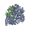
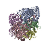
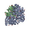
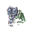
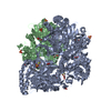
- PDB-2hmo: Crystal Structure of Naphthalene 1,2-Dioxygenase Bound to 3-Nitro... -
+ Open data

- Basic information
| Entry | Database: PDB / ID: 2hmo | ||||||
|---|---|---|---|---|---|---|---|
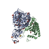
| Title | Crystal Structure of Naphthalene 1,2-Dioxygenase Bound to 3-Nitrotoluene. | ||||||
 Components Components | (Naphthalene 1,2-dioxygenase ...) x 2 | ||||||
 Keywords Keywords | OXIDOREDUCTASE / Protein / Rieske Oxygenase | ||||||
| Function / homology |  Function and homology information Function and homology informationnaphthalene 1,2-dioxygenase / naphthalene 1,2-dioxygenase activity / 3-phenylpropionate catabolic process / dioxygenase activity / 2 iron, 2 sulfur cluster binding / iron ion binding Similarity search - Function | ||||||
| Biological species |  Pseudomonas sp. (bacteria) Pseudomonas sp. (bacteria) | ||||||
| Method |  X-RAY DIFFRACTION / SYNCHROTRON / RIGID BODY REFINEMENT / Resolution: 1.6 Å X-RAY DIFFRACTION / SYNCHROTRON / RIGID BODY REFINEMENT / Resolution: 1.6 Å | ||||||
 Authors Authors | Ferraro, D.J. / Okerlund, A.L. / Mowers, J.C. / Ramaswamy, S. | ||||||
 Citation Citation | Journal: J.Bacteriol. / Year: 2006 Title: Structural basis for regioselectivity and stereoselectivity of product formation by naphthalene 1,2-dioxygenase. Authors: Ferraro, D.J. / Okerlund, A.L. / Mowers, J.C. / Ramaswamy, S. | ||||||
| History |
|
- Structure visualization
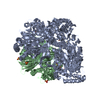
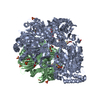
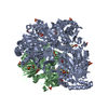
| Structure viewer | Molecule: MolmilJmol/JSmol |
|---|
- Downloads & links
-Download
| PDBx/mmCIF format | 2hmo.cif.gz | 275.3 KB | Display | PDBx/mmCIF format |
|---|---|---|---|---|
| PDB format | pdb2hmo.ent.gz | 219.1 KB | Display | PDB format |
| PDBx/mmJSON format | 2hmo.json.gz | Tree view | PDBx/mmJSON format | |
| Others |  Other downloads Other downloads |
-Validation report
| Arichive directory | https://data.pdbj.org/pub/pdb/validation_reports/hm/2hmoftp://data.pdbj.org/pub/pdb/validation_reports/hm/2hmo | HTTPS FTP |
|---|
-Related structure data
| Related structure data |  2hmjC 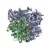 2hmkC  2hmlC 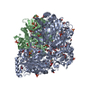 2hmmC  2hmnC 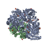 1o7hS S: Starting model for refinement C: citing same article ( |
|---|---|
| Similar structure data |


-Links
 PDBj
PDBj


- Assembly
| Deposited unit | 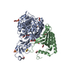
| |||||||||||||||
|---|---|---|---|---|---|---|---|---|---|---|---|---|---|---|---|---|
| 1 | 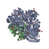
| |||||||||||||||
| Unit cell |
| |||||||||||||||
| Components on special symmetry positions |
| |||||||||||||||
| Details | The biological assembly is and alpha3 beta3 hexamer. One alpha-beta dimer is in the assymetric unit. The others can be generated by the three-fold axis: y, -x-y, z -x-y, x, z |
-Components
-Naphthalene 1,2-dioxygenase ... , 2 types, 2 molecules AB
| #1: Protein | Mass: 49664.355 Da / Num. of mol.: 1 Source method: isolated from a genetically manipulated source Source: (gene. exp.) Pseudomonas sp. (bacteria) / Gene: doxB / Plasmid: pDTG121 / Production host: |
|---|---|
| #2: Protein | Mass: 22969.088 Da / Num. of mol.: 1 Source method: isolated from a genetically manipulated source Source: (gene. exp.) Pseudomonas sp. (bacteria) / Gene: doxD / Plasmid: pDTG121 / Production host: |
-Non-polymers , 6 types, 740 molecules 


| #3: Chemical | ChemComp-FE /  Mass: 55.845 Da / Num. of mol.: 1 / Source method: obtained synthetically / Formula: Fe Mass: 55.845 Da / Num. of mol.: 1 / Source method: obtained synthetically / Formula: Fe | ||||||||
|---|---|---|---|---|---|---|---|---|---|
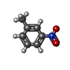
| #4: Chemical | ChemComp-SO4 /  Mass: 96.063 Da / Num. of mol.: 4 / Source method: obtained synthetically / Formula: SO4 Mass: 96.063 Da / Num. of mol.: 4 / Source method: obtained synthetically / Formula: SO4#5: Chemical | ChemComp-FES / |  Mass: 175.820 Da / Num. of mol.: 1 / Source method: obtained synthetically / Formula: Fe2S2 Mass: 175.820 Da / Num. of mol.: 1 / Source method: obtained synthetically / Formula: Fe2S2#6: Chemical | ChemComp-3NT / |  Mass: 137.136 Da / Num. of mol.: 1 / Source method: obtained synthetically / Formula: C7H7NO2 Mass: 137.136 Da / Num. of mol.: 1 / Source method: obtained synthetically / Formula: C7H7NO2#7: Chemical | ChemComp-EDO /  Mass: 62.068 Da / Num. of mol.: 17 / Source method: obtained synthetically / Formula: C2H6O2 Mass: 62.068 Da / Num. of mol.: 17 / Source method: obtained synthetically / Formula: C2H6O2#8: Water | ChemComp-HOH / | Mass: 18.015 Da / Num. of mol.: 716 / Source method: isolated from a natural source / Formula: H2O |
-Experimental details
-Experiment
| Experiment | Method: X-RAY DIFFRACTION / Number of used crystals: 1 |
|---|
- Sample preparation
| Crystal | Density Matthews: 2.71 Å3/Da / Density % sol: 54.64 % |
|---|---|
| Crystal grow | Temperature: 279.15 K / Method: vapor diffusion, hanging drop / pH: 5 Details: 1.9-2.2 M Ammonium Sulfate, 4-6% Dioxane, 0.1 M MES, pH 5.0, VAPOR DIFFUSION, HANGING DROP, temperature 279.15K |
-Data collection
| Diffraction | Mean temperature: 100 K |
|---|---|
| Diffraction source | Source: SYNCHROTRON / Site: ALS  / Beamline: 4.2.2 / Wavelength: 1.04021 Å / Beamline: 4.2.2 / Wavelength: 1.04021 Å |
| Detector | Type: NOIR-1 / Detector: CCD / Date: Nov 3, 2004 |
| Radiation | Monochromator: Double Crystal / Protocol: SINGLE WAVELENGTH / Monochromatic (M) / Laue (L): M / Scattering type: x-ray |
| Radiation wavelength | Wavelength: 1.04021 Å / Relative weight: 1 |
| Reflection | Resolution: 1.6→15.95 Å / Num. all: 102411 / Num. obs: 102411 / % possible obs: 99.2 % / Observed criterion σ(F): 0 / Observed criterion σ(I): 0 / Redundancy: 7.07 % / Rmerge(I) obs: 0.1 / Χ2: 0.95 / Net I/σ(I): 11.8 / Scaling rejects: 5470 |
| Reflection shell | Resolution: 1.6→1.66 Å / Redundancy: 2.62 % / Rmerge(I) obs: 0.43 / Mean I/σ(I) obs: 2.1 / Num. measured all: 25950 / Num. unique all: 9895 / Χ2: 0.86 / % possible all: 96.6 |
- Processing
| Software |
| |||||||||||||||||||||||||||||||||||||||||||||||||||||||||||||||||||||||||||||||||||||||||||||||||||||||||||||||||||||||||||||||||||||||||||||||||
|---|---|---|---|---|---|---|---|---|---|---|---|---|---|---|---|---|---|---|---|---|---|---|---|---|---|---|---|---|---|---|---|---|---|---|---|---|---|---|---|---|---|---|---|---|---|---|---|---|---|---|---|---|---|---|---|---|---|---|---|---|---|---|---|---|---|---|---|---|---|---|---|---|---|---|---|---|---|---|---|---|---|---|---|---|---|---|---|---|---|---|---|---|---|---|---|---|---|---|---|---|---|---|---|---|---|---|---|---|---|---|---|---|---|---|---|---|---|---|---|---|---|---|---|---|---|---|---|---|---|---|---|---|---|---|---|---|---|---|---|---|---|---|---|---|---|---|
| Refinement | Method to determine structure: RIGID BODY REFINEMENT Starting model: 1O7H Resolution: 1.6→15.95 Å / Cor.coef. Fo:Fc: 0.97 / Cor.coef. Fo:Fc free: 0.956 / SU B: 3.048 / SU ML: 0.054 / Cross valid method: THROUGHOUT / σ(F): 0 / ESU R: 0.092 / ESU R Free: 0.079 / Stereochemistry target values: MAXIMUM LIKELIHOOD / Details: HYDROGENS HAVE BEEN ADDED IN THE RIDING POSITIONS
| |||||||||||||||||||||||||||||||||||||||||||||||||||||||||||||||||||||||||||||||||||||||||||||||||||||||||||||||||||||||||||||||||||||||||||||||||
| Solvent computation | Ion probe radii: 0.8 Å / Shrinkage radii: 0.8 Å / VDW probe radii: 1.4 Å / Solvent model: BABINET MODEL WITH MASK | |||||||||||||||||||||||||||||||||||||||||||||||||||||||||||||||||||||||||||||||||||||||||||||||||||||||||||||||||||||||||||||||||||||||||||||||||
| Displacement parameters | Biso mean: 17.437 Å2
| |||||||||||||||||||||||||||||||||||||||||||||||||||||||||||||||||||||||||||||||||||||||||||||||||||||||||||||||||||||||||||||||||||||||||||||||||
| Refinement step | Cycle: LAST / Resolution: 1.6→15.95 Å
| |||||||||||||||||||||||||||||||||||||||||||||||||||||||||||||||||||||||||||||||||||||||||||||||||||||||||||||||||||||||||||||||||||||||||||||||||
| Refine LS restraints |
| |||||||||||||||||||||||||||||||||||||||||||||||||||||||||||||||||||||||||||||||||||||||||||||||||||||||||||||||||||||||||||||||||||||||||||||||||
| LS refinement shell | Resolution: 1.6→1.642 Å / Total num. of bins used: 20
|
